Human S1P1/EDG-1 Antibody
R&D Systems, part of Bio-Techne | Catalog # MAB2016
Clone 218713 was used by HLDA to establish CD designation


Conjugate
Catalog #
Key Product Details
Validated by
Biological Validation
Species Reactivity
Validated:
Human
Cited:
Human, Rat
Applications
Validated:
CyTOF-ready, Flow Cytometry
Cited:
Flow Cytometry, Western Blot
Label
Unconjugated
Antibody Source
Monoclonal Mouse IgG2B Clone # 218713
Product Specifications
Immunogen
NS0 mouse myeloma cell line transfected with human S1P1/EDG‑1
Met1-Ser382
Accession # P21453
Met1-Ser382
Accession # P21453
Specificity
Detects human S1P1/EDG‑1 by flow cytometry.
Clonality
Monoclonal
Host
Mouse
Isotype
IgG2B
Scientific Data Images for Human S1P1/EDG-1 Antibody
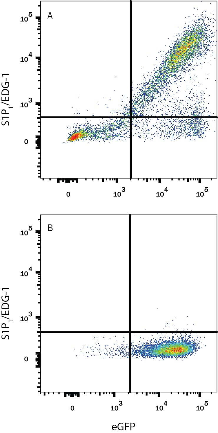
Detection of S1P1/EDG‑1 in CHO Chinese Hamster Cell Line Transfected with Human S1P1/EDG-1 and eGFP by Flow Cytometry.
CHO Chinese hamster ovary cell line transfected with either (A) human S1P1/EDG-1 or (B) irrelevant transfectants and eGFP were stained with Mouse Anti-Human S1P1/EDG-1 Monoclonal Antibody (Catalog # MAB2016) followed by Phycoerythrin-conjugated Anti-Mouse IgG Secondary Antibody (Catalog # F0102B). Quadrant markers were set based on control antibody staining (Catalog # MAB0041). View our protocol for Staining Membrane-associated Proteins.Detection of Human S1P1/EDG-1 by Flow Cytometry
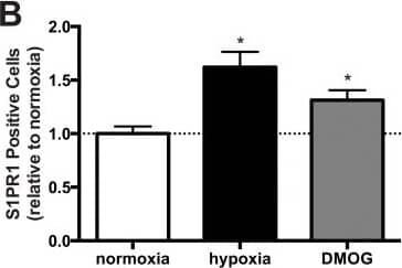
S1PR1 expression by OECs is dynamically altered by microenvironment.OEC expression of S1PR1 under normoxia and hypoxia was qualitatively confirmed by immunocytochemistry (A). The percentage of cells expressing S1PR1 on the cell surface, as confirmed with flow cytometry, was enhanced under hypoxia as compared to the normoxic control (B). Combined stimuli of S1P and VEGF resulted in greater S1PR1 mRNA production after 24h in normoxia (C). Combined stimuli also resulted in a greater proportion of S1PR1+ OECs under both normoxia and hypoxia (D and E). Scale bars represent 40 μm. Data are mean ± SD (n = 4) and normalized to the average untreated control values for either normoxia or hypoxia (C, E; indicated by dashed line) accordingly. An asterisk indicates statistically significant differences (P<0.05) between conditions in normoxia or hypoxia respectively and N.S. displays no statistically significant difference between conditions. Image collected and cropped by CiteAb from the following publication (https://dx.plos.org/10.1371/journal.pone.0123437), licensed under a CC-BY license. Not internally tested by R&D Systems.Applications for Human S1P1/EDG-1 Antibody
Application
Recommended Usage
CyTOF-ready
Ready to be labeled using established conjugation methods. No BSA or other carrier proteins that could interfere with conjugation.
Flow Cytometry
0.25 µg/106 cells
Sample: CHO Chinese hamster ovary cell line transfected with human S1P1/EDG-1 and eGFP
Sample: CHO Chinese hamster ovary cell line transfected with human S1P1/EDG-1 and eGFP
Please Note: Optimal dilutions of this antibody should be experimentally determined.
Reviewed Applications
Read 1 review rated 3 using MAB2016 in the following applications:
Formulation, Preparation, and Storage
Purification
Protein A or G purified from hybridoma culture supernatant
Reconstitution
Reconstitute at 0.5 mg/mL in sterile PBS. For liquid material, refer to CoA for concentration.
Formulation
Lyophilized from a 0.2 μm filtered solution in PBS with Trehalose. *Small pack size (SP) is supplied either lyophilized or as a 0.2 µm filtered solution in PBS.
Shipping
Lyophilized product is shipped at ambient temperature. Liquid small pack size (-SP) is shipped with polar packs. Upon receipt, store immediately at the temperature recommended below.
Stability & Storage
Use a manual defrost freezer and avoid repeated freeze-thaw cycles.
- 12 months from date of receipt, -20 to -70 °C as supplied.
- 1 month, 2 to 8 °C under sterile conditions after reconstitution.
- 6 months, -20 to -70 °C under sterile conditions after reconstitution.
Background: S1P1/EDG-1
Long Name
Endothelial Differentiation Gene 1
Alternate Names
CD363, EDG1, S1PR1
Gene Symbol
S1PR1
UniProt
Additional S1P1/EDG-1 Products
Product Documents for Human S1P1/EDG-1 Antibody
Product Specific Notices for Human S1P1/EDG-1 Antibody
For research use only
Loading...
Loading...
Loading...
Loading...
Loading...
