Breadcrumb
- Home
- Products
- Th17 Cell Kits
- Th17 Cell Kits Cell Culture Products
- CellXVivo Mouse Th17 Cell Differentiation Kit (CDK017)
CellXVivo Mouse Th17 Cell Differentiation Kit
R&D Systems, part of Bio-Techne | Catalog # CDK017

Key Product Details

Summary for CellXVivo Mouse Th17 Cell Differentiation Kit
For the differentiation of Th17 cells from a preparation of CD4+ T cells isolated from mouse splenocytes.
Key Benefits
- Provides optimized reagents needed to induce Th17 differentiation
- Yields ~200 x 106 cells, of which > 40% are Th17 polarized
- Contains high quality bioactive proteins
- Includes straightforward procedures
- Does not require specialized instrumentation
Why Expand Th17 Cells Ex Vivo?
T helper type 17 (Th17) cells are a subset of CD4+ effector T cells that promote cell-mediated immune responses against extracellular bacteria and fungi. Differentiation into the Th17 lineage is promoted by cytokines such as TGF-beta and IL-6, while their survival and expansion are dependent on IL-21 and IL-23. Th17 cells secrete TNF-alpha, IL-6, IL-9, IL-17A, IL-17F, IL-21, and IL-22. Th17 polarized cells are present in low abundance in normal mouse spleen and peripheral blood. In vitro differentiation of mouse Th17 cells from the larger naïve CD4+ T cell population provides increased numbers of Th17 cells to facilitate downstream research.
This kit contains the following reagents for the ex vivo differentiation of mouse Th17 cells.
- Hamster Anti-Mouse CD3
- Rat Anti-Mouse CD28
- Mouse Th17 Reagent 1
- Mouse Th17 Reagent 2
- Mouse Th17 Reagent 3
- Mouse Th17 Reagent 4
- Mouse Th17 Reagent 5
- Reconstitution Buffer 1
- Reconstitution Buffer 2
- Reconstitution Buffer 3
- 20X Wash Buffer
The quantity of the components in the kit is sufficient to differentiate approximately 50x106 naïve CD4+ T cells, and generate ~200 x 106 cells, of which >40% are Th17 polarized cells within your starting CD4+ T cell population.
Note: Results may vary due to strain age and/or the health of the mice used for isolation.
Stability and Storage
Store the unopened kit at ≤-20 °C. Do not use past the kit expiration date. *Provided this is within the expiration date of the kit.
Limitations
- FOR LABORATORY RESEARCH USE ONLY. NOT FOR USE IN DIAGNOSTIC PROCEDURES.
- The safety and efficacy of this product in diagnostic or other clinical uses has not been established.
- This reagent should not be used beyond the expiration date indicated on the label.
- Results may vary due to variations among cells derived from different donors.
T helper type 17 (Th17) cells are involved in the immune response mounted against specific fungi and extracellular bacteria. In mice, Th17 cells develop from naive CD4+ T cells in the presence of TGF-beta and IL-6. These cytokines induce the STAT3-dependent expression of IL-21, IL-23 R, and the transcription factor, ROR gamma t. IL-21 and IL-23 regulate the establishment and clonal expansion of Th17 cells, while ROR gamma t-induced gene expression leads to the secretion of IL-17A, IL-17F, and IL-22. Cytokines secreted by Th17 cells stimulate chemokine secretion by resident cells, leading to the recruitment of neutrophils and macrophages to sites of inflammation. These cells, in turn, produce additional cytokines and proteases that further exacerbate the immune response. In contrast to mouse Th17 differentiation, Th17 polarization in humans requires IL-1 beta, IL-6, IL-21, and IL-23, but seems to be less dependent upon TGF-beta. One other notable difference is that human Th17 cells secrete IL-26, an IL-10 family cytokine without a murine homologue. Cytokines produced by Th17 cells can have both beneficial and pathogenic effects. While they play a central role in eliminating harmful microbes, persistent secretion of Th17 cytokines promotes chronic inflammation and may be involved in the pathogenesis of inflammatory and autoimmune diseases, including rheumatoid arthritis, multiple sclerosis, and inflammatory bowel disorders.
| Species | Mouse |
| Source | N/A |

|
Differentiated Mouse CD4+ Cells Secrete IL-17. Mouse T cells were differentiated for 5 days under Th17 polarization conditions using reagents included in this kit. On day 5, cell culture supernatant was collected and cytokine secretion was determined using the Mouse IL-17 Quantikine®ELISA Kit, the Mouse IFN-γ Quantikine®ELISA Kit, and the Mouse IL-4 Quantikine®ELISA Kit. |

|
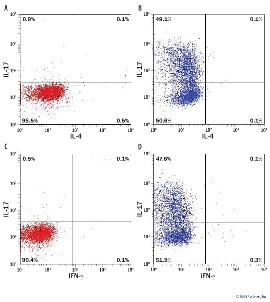
Intracellular Cytokine Staining of Differentiated Mouse Th17 Cells. Flow cytometry data showing mouse naïve CD4+T cells without (A, C) and with (B, D) a 5 day differentiation using reagents included in this kit. On day 5 of differentiation, the cells were stimulated with Cell Activation Cocktail (Tocris®, Catalog # 5476) and stained with Mouse IL-17, Mouse IFN-gamma, and Mouse IL-4 Monoclonal Antibodies. Quadrants were set based on isotype-stained samples. |
Preparation & Storage
| Shipping Conditions | The product is shipped with dry ice or equivalent. Upon receipt, store it immediately at the temperature recommended below. |
| Storage | Store the unopened product at -20 to -70 °C. Use a manual defrost freezer and avoid repeated freeze-thaw cycles. Do not use past expiration date. |
Assay Procedure
Refer to the product datasheet for complete product details.
Briefly, CD4+ T cells isolated from mouse splenocytes are differentiated ex vivo into the Th17 phenotype with reagents provided in the CellXVivo™ Mouse Th17 Cell Differentiation Kit using the following procedure:
- Isolate CD4+ T cells from mouse splenocytes
- Culture CD4+ T cells in reagents provided in kit
- Verify Th17 cell expansion after 5 days in culture
Reagents Supplied in the CellXVivo™ Mouse Th17 Cell Differentiation Kit (Catalog # CDK017):
- Hamster Anti-Mouse CD3
- Rat Anti-Mouse CD28
- Mouse Th17 Reagent 1
- Mouse Th17 Reagent 2
- Mouse Th17 Reagent 3
- Mouse Th17 Reagent 4
- Mouse Th17 Reagent 5
- Reconstitution Buffer 1
- Reconstitution Buffer 2
- Reconstitution Buffer 3
- 20X Wash Buffer
Reagents
- Laboratory mice
- XVIVO™ 15 Chemically Defined, Serum-free Hematopoietic Cell Medium (Lonza, or equivalent)
- MagCellect™ Mouse Naïve CD4+ T Cell Isolation Kit (R&D Systems, Catalog # MAGH115, or equivalent)
- Penicillin/Streptomycin (optional)
- Cell Activation Cocktail 500X (Tocris®, Catalog # 5476)
Equipment
- Tissue culture plates and/or flasks
- Sterile deionized water
- Pipettes and pipette tips
- Inverted microscope
- Hemocytometer
- 37 °C, 5% CO2 incubator
- Centrifuge
Note: Results may vary due to strain, age, and/or the health of the mice used for isolation.
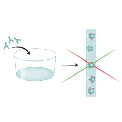
Coat the desired tissue culture plate or flask with Hamster Anti-Mouse CD3 antibody.

Isolate mouse splenocytes.

Perform a cell count.

Perform a cell count.

Suspend 1 x 10⁶ naïve CD4+ T cells/mL in Mouse Th17 Differentiation Media. Culture the cells on plates or flask pre-coated with Hamster Anti-Mouse CD3 antibody.

Refresh with Mouse Th17 Differentiation Media on day 3. Culture for 2 more days.

Verify Th17 cell differentiation by analyzing cytokine expression via flow cytometry or ELISA (optional).

