IL-6 Family Cytokines
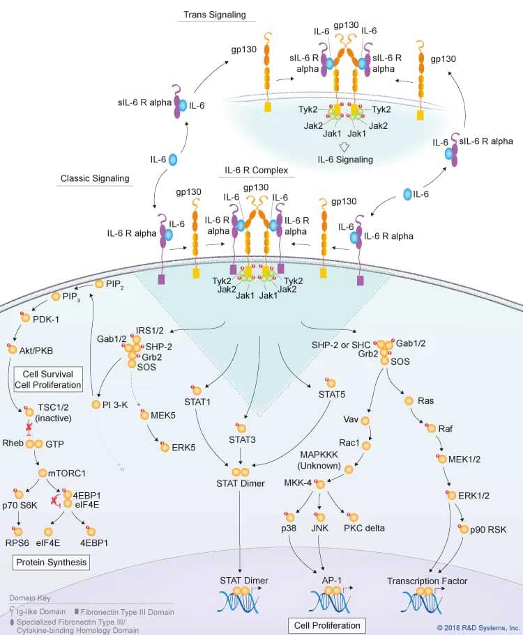
IL-6 Classic and Trans Signaling Pathways
IL-6 Family Cytokines
The IL-6 cytokine family consists of IL-6, IL-11, IL-27 p28/IL-30, IL-31, leukemia inhibitory factor (LIF), oncostatin M (OSM), ciliary neurotrophic factor (CNTF), cardiotrophin-1 (CT-1), cardiotrophin-like cytokine (CLC), and neuropoietin. Although cytokines in this family have pleiotropic functions, they are grouped based on their shared usage of the glycoprotein 130 kDa (gp130) receptor subunit. The exception is IL-31, which uses the gp130-like receptor subunit, IL-31RA. IL-6 family cytokines are also structurally related. With the exception of IL-31 which contains two long and two short chain alpha helices, most IL-6 family cytokines are long chain, four alpha helix bundle cytokines with an up-up-down-down topology. Although members of the IL-6 cytokine family bind to different cytokine-specific receptor subunits, they all recruit the gp130 signaling subunit to form active receptor complexes that promote similar downstream signaling pathways. These pathways include Jak-STAT signaling, Ras-MAPK signaling, PI 3-K-Akt signaling, and p38 and JNK MAPK signaling, which mediate various biological effects in different cell types and result in the unique and overlapping functions of IL-6 family cytokines. Since gp130 is ubiquitously expressed, the more limited expression of the cytokine-specific receptor subunits of the IL-6 family determine which cells are affected by different IL-6 family cytokines. Significantly, a number of cytokines belonging to the IL-6 family including IL-6, IL-11, and CNTF have been shown to be capable of initiating trans-signaling pathways through their interactions with soluble cytokine-specific receptor subunits that are generated either by alternative splicing or by proteolytic cleavage. Formation of these cytokine-soluble receptor complexes can then trans-activate signaling pathways in gp130-expressing cells, thus allowing a wider range of cells to be affected by these cytokines. Read more about select IL-6 family cytokines below.
IL-6
IL-6 is a pleiotropic cytokine that is involved in regulating a wide range of biological processes, including hematopoiesis, the acute phase response, inflammation, liver regeneration, metabolism, bone homeostasis, and cancer progression. It is produced by a variety of immune cell types including dendritic cells, monocytes, and macrophages following toll-like receptor (TLR) activation, along with subsets of activated T cells, B cells, neutrophils, mesenchymal cells, fibroblasts, keratinocytes, astrocytes, and endothelial cells, following treatment with various stimuli. Classic IL-6 signaling is initiated by IL-6 binding to the membrane-bound form of its cytokine-specific receptor subunit, IL-6 R alpha, which is primarily expressed on macrophages, monocytes, dendritic cells, neutrophils, resting lymphocytes, and hepatocytes. The IL-6/IL-6 R alpha complex subsequently associates with gp130, which promotes gp130 dimerization and formation of a 2:2:2 heterohexameric IL-6/IL-6 R alpha/gp130 complex that triggers downstream signaling pathways. IL-6 can also initiate trans-signaling by binding to a soluble form of IL-6 R alpha, which subsequently associates with the membrane-bound gp130 protein on a variety of cell types to activate intracellular signaling. While similar signaling pathways are activated by both the classic and trans-signaling ligand-receptor complexes, the anti-inflammatory and regenerative effects of IL-6 have been suggested to be mediated by classic IL-6 signaling, while IL-6 trans-signaling drives pro-inflammatory responses.
IL-11
IL-11 is produced by multiple cell types including hematopoietic cells, stromal cells, epithelial cells, endothelial cells, osteoblasts, chondrocytes, and neuronal cells. Similar to IL-6, IL-11 can mediate either classic or trans-signaling. The classic signaling pathway is initiated by IL-11 binding to the membrane-bound form of its cytokine-specific receptor subunit, IL-11 R alpha, which subsequently associates with gp130 to form a high affinity receptor complex. Dimerization of gp130 leads to the formation of a 2:2:2 heterohexameric IL-11/IL-11 R alpha/gp130 ligand-receptor complex. IL-11 can also promote trans-signaling in cells that don’t express IL-11 R alpha, by binding to a soluble form of the IL-11 R alpha receptor subunit. This IL-11/sIL-11 R alpha complex subsequently associates with membrane-bound gp130 to form an active receptor complex. IL-11 is another pleiotropic cytokine with a wide range of effects on different cell types. It has been shown to promote the development and maturation of megakaryocytes, stimulate thrombopoiesis and erythropoiesis, regulate neurogenesis, promote osteoclast development, inhibit adipogenesis, regulate epithelial cell proliferation, and have protective effects on endothelial cells. Additionally, it regulates immune cell functions such as macrophage differentiation, Th2 and/or Th17 differentiation, and IgG production by B cells.
IL-31
IL-31 is a structurally unique member of the IL-6 cytokine family that is primarily produced by activated CD4+ T cells, particularly Th2 cells, as well as macrophages, dendritic cells, mast cells, basophils, and eosinophils. Unlike other IL-6 family cytokines, the receptor for IL-31 does not contain gp130. Instead IL-31 signals through a heterodimeric receptor complex consisting of the gp130-like receptor, IL-31RA and OSM R beta. Following its secretion, IL-31 initially binds to IL-31RA and then to OSM R beta, which increases its binding affinity to IL-31RA. While OSM R beta is broadly expressed, IL-31RA is primarily expressed on T cells, dendritic cells, macrophages, basophils, eosinophils, and keratinocytes. IL-31 has been shown to induce the expression of pro-inflammatory cytokines and chemokines, stimulate DRG sensory neurons, and regulate cell proliferation. Many studies suggest that IL-31 is involved in the pathogenesis of pruritic skin disorders, allergic rhinitis, and asthma as elevated levels of IL-31 have been found in patients with these disorders. As a result, researchers are investigating whether antibodies that block IL-31 signaling may have therapeutic potential for treating patients with these diseases.
LIF
Leukemia inhibitory factor (LIF) is produced by a variety of cell types and has a wide range of effects. It was cloned based on its ability to induce differentiation and inhibit proliferation of a mouse myeloid leukemia cell line. It has since been found to be involved in maintaining the pluripotent state of embryonic stem cells, regulating hematopoiesis, supporting successful embryo implantation, regulating bone formation and remodeling, stimulating the development of sensory neurons, and promoting the differentiation of both cardiac smooth muscle cells and adipocytes. LIF signals through a heterodimeric receptor complex consisting of LIF R and gp130. The LIF receptor is widely expressed on embryonic stem cells, monocytes, and macrophages, and is found in many different organ systems, including the liver, uterus, bone, kidney, and central nervous system, consistent with its functions.
Oncostatin M
Oncostatin M (OSM) was originally characterized as a cytokine capable of inhibiting the growth of the A375 human melanoma cell line,. It has since been found to have a broad range of effects on many different cell types. OSM is primarily produced by activated monocytes and macrophages, activated T cells, dendritic cells, neutrophils, and osteoblasts. Unlike other IL-6 family cytokines that initially bind to a cytokine-specific receptor subunit, OSM initially binds to gp130 with low affinity and then recruits either LIF R or OSM R to form an active receptor complex. The gp130/LIF R complex is known as the type I OSM receptor, while the gp130/OSM R complex is known as the type II OSM receptor. Similar to human OSM, rat OSM binds to both receptor complexes, but mouse OSM only binds to the gp130/OSM R complex. OSM is involved in regulating a wide range of biological processes including hematopoiesis, inflammation, bone metabolism, development and homeostasis of the central nervous system, hepatocyte proliferation, the acute phase response, and tissue remodeling during liver regeneration. It has been suggested to be involved in the pathogenesis of several chronic inflammatory conditions including rheumatoid arthritis, inflammatory lung diseases, psoriasis, atopic dermatitis, atherosclerosis, and cardiovascular disease, and seems to play a context-dependent role in different types of cancers.
CNTF
Ciliary neurotrophic factor (CNTF) was originally characterized as a trophic factor for embryonic chick ciliary neurons and was subsequently found to function as a survival factor for embryonic motor neurons, hippocampal neurons, dorsal root ganglion sensory neurons, and sympathetic ganglion neurons. CNTF is primarily expressed by glial cells in the central nervous system and may also be released by damaged cells. CNTF signals through a tripartite receptor complex consisting of either CNTF R alpha, LIF R, and gp130 or IL-6 R alpha, LIF R, and gp130. It initially interacts with either GPI-anchored CNTF R alpha or IL-6 R alpha, which subsequently recruit LIF R and gp130 as signal-transducing receptor subunits. Significantly, CNTF has also been found to initiate trans-signaling by binding to soluble forms of CNTF R alpha or IL-6 R alpha, which then interact with the membrane-bound LIF R-gp130 heterodimeric complex to form an active receptor complex. CNTF not only functions as a neuronal survival factor, but it has also been shown to promote oligodendrocyte survival and astrocyte differentiation. Additionally, it has neuroprotective effects following nervous system injury and in demyelinating neurological diseases, as well as protective and regenerative effects on denervated and intact skeletal muscle.
Bio-Techne offers a wide selection of products for investigating the functions of IL-6 family cytokines including R&D Systems™ bioactive recombinant proteins and single and multianalyte immunoassays for monitoring immune responses. Our catalog also includes antibodies against IL-6 family cytokines, receptors, and intracellular signaling molecules that are validated for one or more of the following applications: blocking/neutralization, Western blot, flow cytometry, and immunohistochemistry.
IL-6 Family Cytokines - Products by Molecule
| Cardiotrophin-1 (CT-1) | Cardiotrophin-like Cytokine (CLC) | Ciliary neurotrophic factor (CNTF) | IL-6 | IL-11 |
| IL-27 p28/IL-30 | IL-31 | Leukemia Inhibitory Factor (LIF) | Neuropoietin (NP) | Oncostatin M (OSM) |
IL-6 Family Receptors - Products by Molecule
| Cytokine-like Factor 1 (CLF-1) | CNTF R alpha | gp130 | IL-6 R alpha | IL-11 R alpha |
| IL-27 R alpha | IL-31RA | LIF R alpha | OSM R beta | Sortilin |
IL-6 Family Intracellular Signaling - Products by Molecule
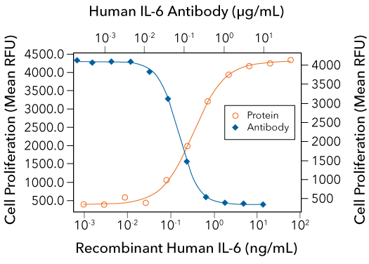
Cell Proliferation Induced by R&D Systems Recombinant Human IL-6 and Neutralization by a Mouse Anti-Human IL-6 Monoclonal Antibody

IL-6-induced Cell Proliferation is Neutralized Using a Mouse Anti-Human IL-6 Monoclonal Antibody. The T1165.85.2.1 mouse plasmacytoma cell line was treated with increasing concentrations of Recombinant Human IL-6 (R&D Systems, Catalog # 206-IL) and cell proliferation was assessed (orange line). The ED50 for this effect is 0.2-0.8 ng/mL. Proliferation stimulated by 2.5 ng/mL Recombinant Human IL-6 was neutralized by treating the cells with increasing concentrations of a Mouse Anti-Human/Primate IL-6 Monoclonal Antibody (R&D Systems, Catalog # MAB206R; blue line). The ND50 for this effect is typically 0.05 - 0.15 ug/mL.
Detection of gp130 in CD3+ Mouse Splenocytes by Flow Cytometry

Detection of gp130 in CD3+ Mouse Splenocytes by Flow Cyotmetry. Mouse splenocytes were stained with an APC-conjugated Rat Anti-Mouse CD3 Monoclonal Antibody (R&D Systems, Catalog # FAB4841A) and either a (A) PE-conjugated Rat Anti-Mouse gp130 Monoclonal Antibody (R&D Systems, Catalog # FAB4681A) or a (B) PE-conjugated Rat IgG2A Isotype Control Antibody (R&D Systems, Catalog # IC006P).
Cell Proliferation Induced by R&D Systems Recombinant Human IL-11 and Neutralization by a Mouse Anti-Human IL-11 Monoclonal Antibody

IL-11-induced Cell Proliferation is Neutralized Using a Mouse Anti-Human IL-11 Monoclonal Antibody. The T11 mouse plasmacytoma cell line was treated with increasing concentrations of Recombinant Human IL-11 (R&D Systems, Catalog # 218-IL) and cell proliferation was assessed (orange line). The ED50 for this effect is 0.02-0.12 ng/mL. Proliferation stimulated by 1 ng/mL Recombinant Human IL-11 was neutralized by treating the cells with increasing concentrations of a Mouse Anti-Human IL-11 Monoclonal Antibody (R&D Systems, Catalog # MAB218; blue line). The ND50 for this effect is typically < 8 ug/mL.
Detection of IL-31RA in IFN-gamma-treated CD14+ Human PBMCs by Flow Cytometry

Detection of IL-31RA in IFN-gamma-treated Human PBMCs by Flow Cyotmetry. Human peripheral blood mononuclear cells (PBMCs) were (A) treated with 50 ng/mL Recombinant Human IFN-gamma (R&D Systems, Catalog # 285-IF) for 20 hours or (B) untreated, and then stained with an APC-conjugated Mouse Anti-Human CD14+ Monoclonal Antibody (R&D Systems, Catalog # FAB3832A) and a Rat Anti-Human IL-31 RA Monoclonal Antibody (R&D Systems, Catalog # MAB2769) followed by a PE-conjugated Goat Anti-Rat IgG Secondary Antibody (R&D Systems, Catalog # F0105B). Quadrant markers were set based on staining with a Rat IgG1 Isotype Control (R&D Systems, Catalog # MAB005).
Cell Proliferation Induced by R&D Systems Recombinant Human LIF and Neutralization by a Goat Anti-Human LIF R alpha Polyclonal Antibody

LIF-induced Cell Proliferation is Neutralized Using a Goat Anti-Human LIF R alpha Polyclonal Antibody. The TF-1 human erythroleukemic cell line was treated with increasing concentrations of Recombinant Human LIF (R&D Systems, Catalog # 7734-LF) and cell proliferation was assessed (orange line). The ED50 for this effect is 0.02-0.12 ng/mL. Proliferation stimulated by 0.3 ng/mL Recombinant Human LIF was neutralized by treating the cells with increasing concentrations of a Goat Anti-Human LIF R alpha Affinity-purified Polyclonal Antibody (R&D Systems, Catalog # AF-249-NA; blue line). The ND50 for this effect is typically 6-36 ug/mL.
Cell Proliferation Induced by R&D Systems Recombinant Mouse Oncostatin M and Neutralization by a Rat Anti-Mouse Oncostatin M Monoclonal Antibody

Oncostatin M-induced Cell Proliferation is Neutralized Using a Rat Anti-Mouse Oncostatin M Monoclonal Antibody. The NIH-3T3 mouse embryonic fibroblast cell line was treated with increasing concentrations of Recombinant Mouse Oncostatin M (R&D Systems, Catalog # 495-MO) and cell proliferation was assessed (orange line). The ED50 for this effect is 0.25-1 ng/mL. Proliferation stimulated by 1 ng/mL Recombinant Mouse Oncostatin M was neutralized by treating the cells with increasing concentrations of a Rat Anti-Mouse Oncostatin M Monoclonal Antibody (R&D Systems, Catalog # MAB4951; blue line). The ND50 for this effect is typically 0.2-1.2 ug/mL.
Immunostaining of CNTF in Perfusion-fixed Frozen Sections of Rat Cerebellum Using R&D Systems Goat Anti-Rat CNTF Polyclonal Antibody

Detection of CNTF in Rat Cerebellum. Perfusion-fixed frozen sections of rat cerebellum were stained for CNTF using a Goat Anti-Mouse/Rat CNTF Antigen Affinity-purified Polyclonal Antibody (R&D Systems, Catalog # AF-557-NA) at 5 ug/mL for 1 hour at room temperature followed by incubation with the Anti-Goat IgG VisUCyte HRP Polymer Antibody (R&D Systems, Catalog # VC004). Tissue was stained using DAB (brown) and counterstained with hematoxylin (blue). Specific CNTF staining was localized to the Purkinje neurons.
Featured Products for IL-6 Family Research

Immunoassays for IL-6 Family Cytokine Detection
Immunoassays for IL-6 Family Cytokine Detection
From our complete, ready-to-use Quantikine ELISA Kits to our more flexible DuoSet ELISA Development Systems, we offer a wide selection of immunoassays for measuring IL-6 family cytokines. Whatever your needs, you can count on our immunoassays to deliver accurate, reproducible, high-quality data for every experimental sample that you test.

Custom Protein Libraries
Custom Protein Libraries
Are you spending valuable research time aliquoting proteins into plates? Let us help. We offer Custom Protein Libraries, which come with the proteins that you select already aliquoted into the wells of a 96-well plate. This custom product allows you to select the proteins that you need from a portfolio of over 5,000 R&D Systems bioactive proteins and will significantly reduce the amount of time needed to set-up your experiments.
Featured IL-6 Family Resources

Periodic Table of Cytokine & Chemokine Families Wall Poster
Periodic Table of Cytokine & Chemokine Families Wall Poster
Learn about the members of different cytokine and chemokine families with our periodic table wall poster. This poster includes information about the structures, receptors, and native molecular weights of each cytokine, and will serve as a great reference tool and colorful addition to your lab space.

Cytokine Signaling Pathways
Cytokine Signaling Pathways
Cytokines activate a diverse array of intracellular signaling pathways that can induce processes such as cell proliferation, differentiation, migration, and inflammation. Explore the signaling pathways that are activated by different cytokine families, the primary target cells that they affect, and the biological effects that they mediate using our interactive signaling pathways.


