By Jennifer Sokolowski, MD, PhD.
Microglia are a major immune-cell component in the brain. They ingest and degrade dead cells, debris, and foreign material and interact with other immune cells to orchestrate central nervous system immune responses.1,2 Microglia appear to play a critical role in modulating normal physiologic immune functions as well as the immune response in disease states. In order to define the role of microglia, we need specific markers that allow distinction of microglia from other cell types; fortunately, researchers have validated a new microglia-specific marker, TMEM 119.3
Microglia: the "resident macrophages" of the brain
Microglia have been referred to as the 'resident macrophages' of the CNS, and because they share such similar characteristics, it is difficult to delineate microglia from peripherally-derived macrophages. Macrophages are myeloid lineage cells which can be replenished by circulating monocytes. Microglia were initially thought to derive from the same precursors; however, studies have shown that microglia actually derive from yolk sac progenitors and migrate early in development to set up residence in the brain parenchyma, and are a self-renewing population.4,5 Commonly used antibody markers to highlight microglia include AIF-1/iba1, F4/80, CX3CR1, CD11b, and CD68, but these are also expressed by macrophages.

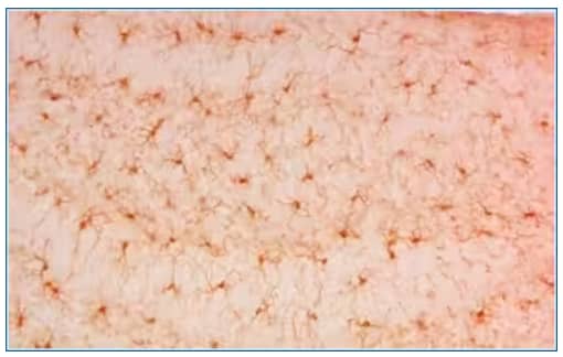
Immunohistochemistry-Paraffin: AIF-1/Iba1 Antibody [NBP2-19019] - Iba1 detects Iba1 protein at cytosol on mouse brain tissue by immunohistochemical analysis. Sample: Paraffin-embedded mouse brain tissue. Iba1 dilution: 1:1000.
Utility of TMEM119 as a marker that distinguishes microglia from macrophages
A 2016 study by Bennet et al. used GeneChip data to identify TMEM 119 as a specific marker of microglia cells and validated this with subsequent RNA in situ hybridization, antibody staining, and quantitative PCR. They subsequently used TMEM119 as a tool to sort microglia and performed transcriptome analysis. They were able to use this approach to characterize RNAseq profiles of highly pure mouse microglia during development and after an immune challenge.3
Markers that highlight microglia and macrophages:
- AIF-1/iba1, ionized calcium binding adaptor molecule 1, is a calcium binding protein that is involved in membrane reorganization
- F4/80, glycoprotein found on the cell surface
- CX3CR1, the fractalkine receptor, responds to fractalkine (aka CX3CL1)
- CD11b, an integrin family member that plays a role in cell adhesion
- CD68, a lysosomal protein
Marker specific for microglia:
- TMEM 119, transmembrane protein 119, a cell-surface protein of unknown function.
The capacity to specifically identify microglia for immunostaining, subsequent cell isolation, transcriptomics and proteomics analysis will help progress our understanding of microglial function in health and disease.
Jennifer Sokolowski, MD, PhD
University of Virginia, Department of Neurosurgery
-
Lopez-Atalaya JP et al. (2018) Development and Maintenance of the Brain’s Immune Toolkit: Microglia and Non-Parenchymal Brain Macrophages Dev Neurobiol 78:561-579.
-
Li, Q et al. (2018) Microglia and macrophages in brain homeostasis and disease. Nat Rev Immunol. 18:225-242.
-
Bennett ML et al. (2016) New tools for studying microglia in the mouse and human CNS Proc Natl Acad Sci USA
-
Ginhoux F et al. (2010) Fate mapping analysis reveals that adult microglia derive from primitive macrophages Science 330:841-845.
-
Gomez Perdiguero, E et al. (2015) Tissue-resident macrophages originate from yolk-sac-derived erythro-myeloid progenitors Nature 518:547-551.