Signal amplification methods are widely used in immunohistochemistry (IHC) for detection of rare epitopes and low abundance antigens. While many of these techniques such as the avidin-biotin complex (ABC) method improve staining, they frequently require additional steps and result in higher background staining. Blocking endogenous biotin, a requirement of using ABC reagents, may not sufficiently remove residual activity in frozen tissue sections and tissues high in biotin including the liver and kidney.
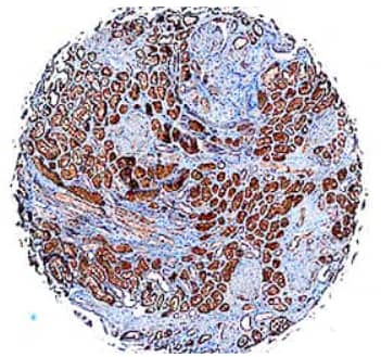
Figure 1. FABP1/L-FABP was detected in paraffin-embedded sections of human kidney using Mouse Anti-Human FABP1/L-FABP Monoclonal Antibody (Catalog # MAB2964) at 1 μg/mL for 1 hour at room temperature followed by incubation for 30 minutes at room temperature with Anti-Mouse/Rabbit IgG VisUCyte HRP Polymer Antibody (Catalog # VC002). Tissue was stained with DAB (brown) and counterstained with hematoxylin (blue).
VisUCyte HRP polymer conjugated secondary antibodies are biotin-free reagents that decrease the number of steps in the IHC staining procedure without compromising the quality of antigen detection (see Figure 1). These secondary antibodies link numerous horseradish peroxidase (HRP) molecules on a polymer backbone to the site of primary antibody binding and increase assay sensitivity by 2-3 fold compared to the ABC method. With the polymer based method there’s no need to block endogenous biotin or to include additional layers for signal amplification. HRP-polymers are directly conjugated to secondary antibodies, enabling a two-step detection process.
In our side-by-side comparison of ABC reagents and HRP polymer secondary antibodies using a fixed concentration of primary antibody, the HRP-polymer conjugates demonstrated a dramatic boost in antigen-specific staining of formalin-fixed, paraffin-embedded (FFPE) tissue sections. To achieve equivalent staining by IHC, the polymer-based method required less primary antibody and a shortened incubation time. The polymer-based amplification method is faster, with less steps, which helps to minimize the complexity of IHC experiments.
VisUCyte HRP polymer secondary antibodies are a great addition to our strong portfolio of IHC detection reagents and address the key limitations of streptavidin-biotin based detection methods. To view data generated using these secondary antibodies and to learn more about the benefits of using them, read our technology brief.