Analyzing Formalin-Fixed, Paraffin-Embedded Tissue on Simple Western
Introduction
Formalin-fixed, paraffin-embedded (FFPE) tissue is a staple of biological and biomedical research. Since the late 19th century, FFPE samples have routinely been prepared during biopsies and autopsies from healthy and diseased tissues.1 Because FFPE samples are stable at room temperature for decades, they are ideal for retrospective analysis to hunt for novel biomarkers implicated in disease. Huge repositories of FFPE samples have been collected worldwide at universities and hospitals. Thus, they are a rich resource to study cancer and other diseases.
Extracting proteins from FFPE samples for proteomic studies is a challenge. Traditional gel-based and Simple Western™ procedures require full-length proteins, while mass spectrometry analysis can be done with intact or digested proteins.2 Efficiently extracting full-length proteins is labor-intensive, and protein yields are often low. This challenge is compounded by the fact that biopsy samples are often small, and sacrificing some of the tissue for proteomic analysis can seriously compromise pathological diagnosis. Despite these challenges, the Qproteome® FFPE Tissue Kit from QIAGEN is a useful method to extract full-length proteins.
Once extracted, the ideal method for FFPE protein analysis combines excellent sensitivity with small sample volume needs. Simple Western is a great solution for studying proteins in FFPE because the sensitivity it provides enables detection of small amounts of protein in your sample, so that even low-yield protein extractions can provide valuable information. Another major advantage is that only small sample sizes are required, thereby preserving precious FFPE samples. Also, protein separation and detection on Simple Western is fully automated, which reduces the complexity of your FFPE workflow, and results are generated in as little as three hours.
Here, we developed a straightforward protocol to extract and analyze FFPE samples by Simple Western. Full-length proteins were extracted with the QIAGEN kit from healthy and cancerous breast tissue. Then, commercial antibodies were used to detect breast cancer biomarkers and other host proteins. We show that minimal FFPE tissue is required for efficient protein detection, so you can get the most information out of your precious FFPE sample.
Materials
ProteinSimple - A Bio-Techne Brand
- Instrument: Jess™, Abby™, Wes™, Peggy Sue™, or Sally Sue™,
- 12–230 kDa Separation Module for Jess, Abby, or Wes (PN SM-W004)
- 12–230 kDa Separation Module for Peggy Sue or Sally Sue (PN SM-S001)
- Anti-Rabbit Detection Module for Jess, Wes, Peggy Sue or Sally Sue (PN DM-001)
- Total Protein Detection Module for Jess, Wes, Peggy Sue or Sally Sue (PN DM-TP01)
Other reagents
- Qproteome FFPE Tissue Kit (QIAGEN, PN 37623)
Samples and antibodies
- 5 microtome sections of healthy and cancerous FFPE tissue samples, each 10-15 mm long and 10 µm thick (Acepix Biosciences)
| Antibody | Supplier | Part Number | Dilution |
|---|---|---|---|
| AKT1 | Cell Signaling Technology | 2938 | 1:50 |
| ERK1/2 | ProteinSimple, a Bio-Techne Brand | 040-474 | Ready-To-Use |
| Flotillin-1 | Novus Biologicals, a Bio-Techne Brand | NBP2-16508 | 1:20 |
| GAPDH | Cell Signaling Technology | 2118 | 1:50 |
| HER2 | Cell Signaling Technology | 4290 | 1:25 |
| HSP60 | R&D Systems, a Bio-Techne Brand | AF1800 | 1:50 |
| pERK1/2 | Cell Signaling Technology | 4370 | 1:50 |
| SRC | Cell Signaling Technology | 2107 | 1:50 |
Table 1. Primary antibodies used in this study, all sourced from rabbit.
Methods
- Follow the instructions in the QIAGEN Qproteome FFPE Tissue Handbook to extract full-length proteins from your FFPE samples. The handbook also provides protein yields from different tissues. We recommend aliquoting the processed samples and placing them at -20 °C or -80 °C for long-term storage.
- Prepare 0.1X Sample Buffer by diluting the 10X Sample Buffer 1:100 in DI water.
- To identify the linear range of detection of your samples, create a serial dilution series in 0.1X Sample Buffer. We recommend starting with a 2-fold dilution series, for example 1:2, 1:4, 1:8, etc.
- Dilute the antibodies in Antibody Diluent 2.
- Follow the default sample preparation and assay conditions for Simple Western. You can use the Simple Western Total Protein Assay to normalize your immunoassay data.
FFPE Tissue Sample Results
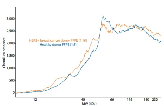
The total protein in the healthy and diseased samples was assessed by the Simple Western Total Protein Assay. A 2-fold serial dilution series of each sample was created and loaded on Simple Western. This analysis revealed that the extract from the diseased sample contained about twice as much protein as the extract from the healthy sample (Figure 1).
A major advantage of the Simple Western Total Protein Assay for FFPE applications is that it bypasses interference of the reducing agent β-mercaptoethanol (β-ME) that is present in processed FFPE extracts. β-ME is well known to interfere with common total protein assays, such as the BCA assay.
With this information, we were able to load equivalent total protein of each sample in our analysis to accurately compare relative expression of target proteins between the healthy and diseased samples. We tested expression of several intracellular targets, including AKT, ERK1/2, Flotillin-1, GAPDH, HSP60 and SRC (Figure 2). All of these targets were successfully detected, and most of these proteins showed an increase in expression in the tumorous tissue (Figure 2), indicating increased metabolic activity and cellular proliferation that is characteristic of tumor growth.3
Because the expression of cell surface receptor HER2 is a hallmark of this particular breast cancer sample type, we tested whether we could detect an increase in expression of HER2 in diseased tissue compared to healthy tissue. As expected, a strong signal for HER2 was detected in breast cancer tissue over the healthy donor tissue (Figure 3).

Figure 1. Overlaid electropherograms of healthy and diseased FFPE samples as measured by the Simple Western Total Protein Assay. The total protein in the two samples was approximately equivalent when the healthy sample was diluted 1:5 and the diseased sample was diluted 1:10.

Figure 2. (A) Electropherogram of phosphorylated ERK1/2 (pERK1/2) in healthy and breast cancer donor FFPE samples. (B) Lane view of targets detected in healthy (H) and diseased (D) FFPE extracts. To load equivalent total protein samples, the healthy extract was diluted 1:15 and the diseased extract was diluted 1:30.

Figure 3. (A) Lane view of HER2 expression in healthy (H) and diseased (D) FFPE extracts. (B) Overlaid electropherograms of HER2 detected in diseased and healthy FFPE extracts. To load equivalent total protein samples, the healthy extract was diluted 1:15 and the diseased extract was diluted 1:30. The noisy HER2 separation profile shown here is typical for an FFPE extraction, as was shown previously by traditional Western blot analysis.4
Conclusion
Simple Western is a great solution for proteomic analysis of FFPE tissue samples. Only a small amount of FFPE extract is required; as little as 0.5 µm3 of a microtome section could be analyzed per well on Simple Western, and at least 500 Simple Western samples could be prepared from a single FFPE extraction. You can also analyze up to 25 samples with Jess or Abby, and up to 96 samples with Sally Sue or Peggy Sue, all in a fully automated fashion and with a built-in total protein assay that is unaffected by reducing agents like β-ME.
-
Fox et al. (1985) Formaldehyde fixation Journal of Histochemistry & Cytochemistry 33:845-53. PMID: 3894502.
-
Gustafsson et al. (2015) Proteomic developments in the analysis of formalin-fixed tissue Biochimica et Biophysica Acta (BBA) - Proteins and Proteomics 1854:559-80. PMID: 25315853.
-
Romero-Garcia et al. (2011) Tumor cell metabolism: an integral view 1:939-48. PMID: 22057267.
-
Becker et al. (2008) Extraction of Phosphorylated Proteins from Formalin-Fixed Cancer Cells and Tissues The Open Pathology Journal 2:46-52.