8-oxo-dG Antibody
R&D Systems, part of Bio-Techne | Catalog # 4354-MC-050

Key Product Details
Species Reactivity
Validated:
Multi-Species
Cited:
Human, Mouse, Rat, Insect - Drosophila, Transgenic Mouse
Applications
Validated:
ELISA, Immunocytochemistry, Immunohistochemistry
Cited:
Bioassay, Chromatin Immunoprecipitation (ChIP), Co-Immunoprecipitation, Comet Assay, Immunocytochemistry, Immunohistochemistry, Immunohistochemistry-Frozen, Western Blot
Label
Unconjugated
Antibody Source
Monoclonal Mouse IgG2B Clone # 15A3
Product Specifications
Immunogen
8-oxo-dG-conjugated-KLH
Specificity
This mouse monoclonal antibody specifically binds to 8-hydroxy-2'-
deoxyguanosine within DNA in H2O2-treated cells.
Clonality
Monoclonal
Host
Mouse
Isotype
IgG2B
Scientific Data Images for 8-oxo-dG Antibody
Detection of Rat 8-oxo-dG 8-oxo-dG Antibody (15A3) by Immunohistochemistry
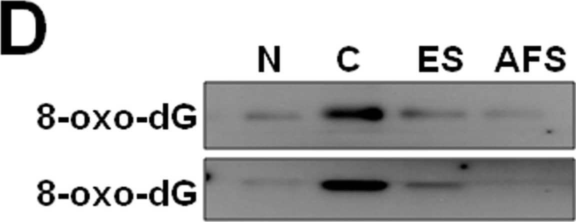
Expression of apoptotic factors in denervated muscle 2 weeks after denervation.(A) Expression of Bcl-2, Bad, and Bax in denervated muscle subjected to different treatment (B) Quantitative analysis of apoptotic markers in different treatment groups (C) The expression of 8-oxo-dG in normal, control, ES, and AFS group. (D) The representative of western blot analysis of 8-oxo-dG related to different treatment (n = 2) (F) Quantitative analysis of caspase-3 in different treatment groups Bar length = 100 μm. R = right, L = left, N = 6 for each group, * p<0.05 and **p<0.01indicated a statistical difference compared to the control group; # p<0.05 indicated a statistical difference as compared to the ES group. Image collected and cropped by CiteAb from the following publication (https://pubmed.ncbi.nlm.nih.gov/25945496), licensed under a CC-BY license. Not internally tested by R&D Systems.Detection of Rat 8-oxo-dG 8-oxo-dG Antibody (15A3) by Western Blot
Expression of apoptotic factors in denervated muscle 2 weeks after denervation.(A) Expression of Bcl-2, Bad, and Bax in denervated muscle subjected to different treatment (B) Quantitative analysis of apoptotic markers in different treatment groups (C) The expression of 8-oxo-dG in normal, control, ES, and AFS group. (D) The representative of western blot analysis of 8-oxo-dG related to different treatment (n = 2) (F) Quantitative analysis of caspase-3 in different treatment groups Bar length = 100 μm. R = right, L = left, N = 6 for each group, * p<0.05 and **p<0.01indicated a statistical difference compared to the control group; # p<0.05 indicated a statistical difference as compared to the ES group. Image collected and cropped by CiteAb from the following publication (https://pubmed.ncbi.nlm.nih.gov/25945496), licensed under a CC-BY license. Not internally tested by R&D Systems.Applications for 8-oxo-dG Antibody
Application
Recommended Usage
ELISA
Empirical determination will be required for optimal results.
Immunocytochemistry
1:250 dilution
Sample:
Sample:
H2O2 treated MCF 10A human breast epithelial cell line
Immunohistochemistry
1:250 dilution
Sample: Paraffin embedded rat thymus tissue
Sample: Paraffin embedded rat thymus tissue
Reviewed Applications
Read 4 reviews rated 4.8 using 4354-MC-050 in the following applications:
Formulation, Preparation, and Storage
Purification
Protein A or G purified from ascites
Formulation
This antibody is provided as purified immunoglobulin from mouse ascites
at 0.5 mg/ml in 1X PBS containing 0.1% sodium azide, 50% glycerol.
Shipping
The product is shipped with dry ice or equivalent. Upon receipt, store it immediately at the temperature recommended below.
Stability & Storage
Store the unopened product at -20 to -70 °C. Use a manual defrost freezer and avoid repeated freeze-thaw cycles. Do not use past expiration date.
Background: 8-oxo-dG
Long Name
8-Hydroxyguanine
Alternate Names
8-OHG, 8oxodG
Additional 8-oxo-dG Products
Product Documents for 8-oxo-dG Antibody
Product Specific Notices for 8-oxo-dG Antibody
For research use only
Loading...
Loading...
Loading...
Loading...

