Caspase-3 Antibody (31A1067) - (Pro and Active) - Azide and BSA Free
Novus Biologicals, part of Bio-Techne | Catalog # NBP2-33244

Conjugate
Catalog #
Forumulation
Catalog #
Key Product Details
Validated by
Biological Validation
Species Reactivity
Validated:
Human, Mouse, Rat, Porcine, Chicken, Chinese Hamster, Mammal
Cited:
Human, Mouse
Applications
Flow Cytometry, Immunocytochemistry/ Immunofluorescence, Immunohistochemistry, Immunohistochemistry-Frozen, Immunohistochemistry-Paraffin, Knockdown Validated, Knockout Validated, Simple Western, Western Blot
Label
Unconjugated
Antibody Source
Monoclonal Mouse IgG1 kappa Clone # 31A1067
Format
Azide and BSA Free
Concentration
Please see the vial label for concentration. If unlisted please contact technical services.
Product Specifications
Immunogen
This Caspase-3 Antibody (31A1067) - (Pro and Active) - Azide and BSA Free was developed against full-length recombinant human caspase-3 protein.
Specificity
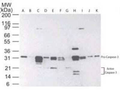
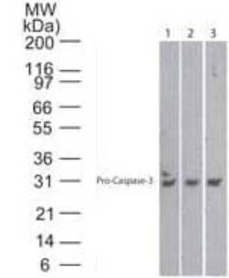
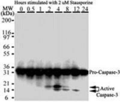
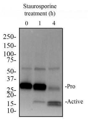
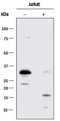
The antibody detects both pro Caspase-3 (~32 kDa) and the large subunit of the active/cleaved form (~14-21 kDa) of Caspase-3.
Clonality
Monoclonal
Host
Mouse
Isotype
IgG1 kappa
Theoretical MW
31.7 kDa.
Disclaimer note: The observed molecular weight of the protein may vary from the listed predicted molecular weight due to post translational modifications, post translation cleavages, relative charges, and other experimental factors.
Disclaimer note: The observed molecular weight of the protein may vary from the listed predicted molecular weight due to post translational modifications, post translation cleavages, relative charges, and other experimental factors.
Scientific Data Images for Caspase-3 Antibody (31A1067) - (Pro and Active) - Azide and BSA Free
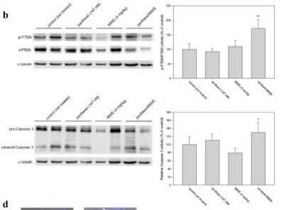
Western Blot Analysis of Caspase-3 in TSGH 8301 Cells After Multiple Treatment Types
Paclitaxel in combination with MWE retarded tumor growth in a human bladder carcinoma TSGH 8301 xenograft model. The levels of total (t-PTEN) and phospho-PTEN (p-PTEN) and Caspase 3 in the tumor specimens were determined by Western blotting and then quantified using beta-actin as the protein loading control; the results are expressed as a percentage of the control. Image collected and cropped by CiteAb from the following publication (https://www.nature.com/articles/srep20417), licensed under a CC-BY license. Image using the standard format of this product.Immunohistochemical Staining of Caspase-3 in Paraffin Embedded Human Spleen
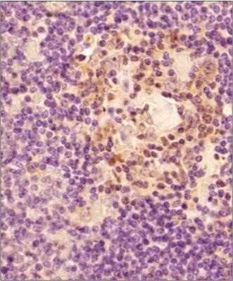
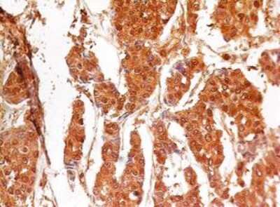
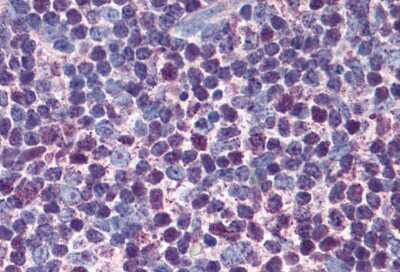
Tissue section of human spleen using 1:200 dilution of Caspase-3 antibody (clone 31A1067). The staining was developed with HRP labeled anti-mouse IgG secondary antibody and DAB reagent, and nuclei of cells were counter-stained with hematoxylin. This Caspase 3 antibody generated primarily a specific cytoplasmic staining in a subset of spleenocytes with some nuclear signal in a few cells. Image using the standard format of this product.Flow Cytometry of HeLa Cells Stained with Caspase-3 Antibody
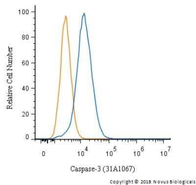
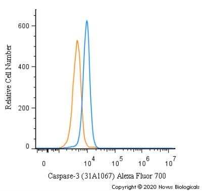
An intracellular stain was performed on HeLa cells with Caspase-3 Antibody (31A1067) - (Pro and Active) NB100-56708 (blue) and a matched isotype control (orange). Cells were fixed with 4% PFA and then permeablized with 0.1% saponin. Cells were incubated in an antibody dilution of 5 ug/mL for 30 minutes at room temperature, followed by mouse F(ab)2 IgG (H+L) APC-conjugated secondary antibody (F0101B, R&D Systems). Image using the standard format of this product.Applications for Caspase-3 Antibody (31A1067) - (Pro and Active) - Azide and BSA Free
Application
Recommended Usage
Immunocytochemistry/ Immunofluorescence
reported in scientific literature (PMID 23840553)
Immunohistochemistry
1:10-1:500
Immunohistochemistry-Frozen
1:10-1:500. Use reported in scientific literature (Zhang et al)
Immunohistochemistry-Paraffin
1:10-1:500. Use reported in scientific literature (Lee et al)
Western Blot
1-5 ug/ml
Application Notes
The large subunit of the cleaved form may appear as one or two or even as a stack of bands depending on the presence or absence of the Caspase-3 pro-domain. It is highly recommended that a maximum sensitivity ECL substrate (Femto sensitive) be used for efficient detection of this antibody in Western blot applications.
Formulation, Preparation, and Storage
Purification
Protein G purified
Formulation
PBS - BSA free
Format
Azide and BSA Free
Preservative
No Preservative
Concentration
Please see the vial label for concentration. If unlisted please contact technical services.
Shipping
The product is shipped with polar packs. Upon receipt, store it immediately at the temperature recommended below.
Stability & Storage
Store at 4C short term. Aliquot and store at -20C long term. Avoid freeze-thaw cycles.
Background: Caspase-3
References
1.Mu, N., Lei, Y., Wang, Y., Wang, Y., Duan, Q., Ma, G., . . . Su, L. (2019). Inhibition of SIRT1/2 upregulates HSPA5 acetylation and induces pro-survival autophagy via ATF4-DDIT4-mTORC1 axis in human lung cancer cells. Apoptosis, 24(9-10), 798-811. doi:10.1007/s10495-019-01559-3
2.Sun, C. M., Enkhjargal, B., Reis, C., Zhou, K. R., Xie, Z. Y., Wu, L. Y., . . . Zhang, J. H. (2019). Osteopontin attenuates early brain injury through regulating autophagy-apoptosis interaction after subarachnoid hemorrhage in rats. CNS Neurosci Ther, 25(10), 1162-1172. doi:10.1111/cns.13199
3.Louneva, N., Cohen, J. W., Han, L. Y., Talbot, K., Wilson, R. S., Bennett, D. A., . . . Arnold, S. E. (2008). Caspase-3 is enriched in postsynaptic densities and increased in Alzheimer's disease. Am J Pathol, 173(5), 1488-1495. doi:10.2353/ajpath.2008.080434
Alternate Names
Apopain, CASP3, Caspase3, CPP32, LICE-1, YAMA
Gene Symbol
CASP3
Additional Caspase-3 Products
Product Documents for Caspase-3 Antibody (31A1067) - (Pro and Active) - Azide and BSA Free
Product Specific Notices for Caspase-3 Antibody (31A1067) - (Pro and Active) - Azide and BSA Free
This product is for research use only and is not approved for use in humans or in clinical diagnosis. Primary Antibodies are guaranteed for 1 year from date of receipt.
Loading...
Loading...
Loading...
Loading...
Loading...
