CD34 Antibody (MEC 14.7) - BSA Free
Novus Biologicals, part of Bio-Techne | Catalog # NB600-1071


Conjugate
Catalog #
Forumulation
Catalog #
Key Product Details
Species Reactivity
Validated:
Mouse, Human (Negative)
Cited:
Human, Mouse, Rat, Rabbit
Applications
Validated:
ELISA, Flow Cytometry, Immunocytochemistry/ Immunofluorescence, Immunohistochemistry, Immunohistochemistry-Frozen, Immunohistochemistry-Paraffin, Immunoprecipitation, Multiplex Immunofluorescence, Western Blot
Cited:
Flow Cytometry, IF/IHC, Immunocytochemistry/ Immunofluorescence, Immunohistochemistry-Frozen, Immunohistochemistry-Paraffin, Western Blot
Label
Unconjugated
Antibody Source
Monoclonal Rat IgG2A Clone # MEC 14.7
Format
BSA Free
Concentration
1 mg/ml
Product Specifications
Immunogen
Murine transformed endothelioma cell line t-end.
Reactivity Notes
This antibody does not detect Human CD34 and is an excellent tool for marking host derived endothelial cells/vasculature in human cancer xenografts on mouse.
Localization
Membrane; Single-pass type I membrane protein.
Marker
Hematopoietic Stem Cell Marker
Clonality
Monoclonal
Host
Rat
Isotype
IgG2A
Scientific Data Images for CD34 Antibody (MEC 14.7) - BSA Free
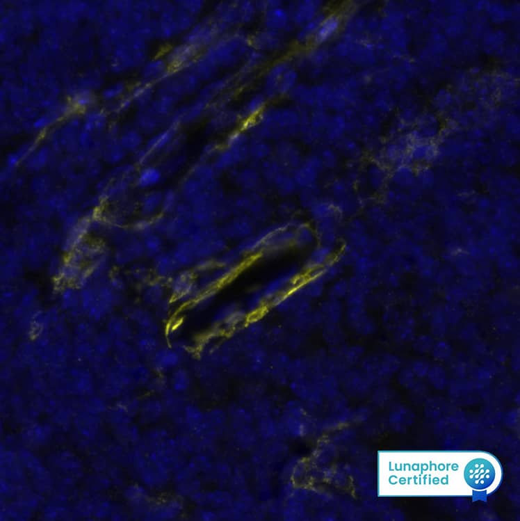
Detection of CD34 in Mouse Thymus via seqIF™ staining on COMET™
CD34 was detected in immersion fixed paraffin-embedded sections of mouse Thymus using Rat Anti-Mouse CD34, Monoclonal Antibody (Catalog #NB600-1071) at 1:500 dilution at 37 ° Celsius for 2 minutes. Before incubation with the primary antibody, tissue underwent an all-in-one dewaxing and antigen retrieval preprocessing using PreTreatment Module (PT Module) and Dewax and HIER Buffer H (pH 9; Epredia Catalog # TA-999-DHBH).Tissue was stained using the Alexa Fluor™ 647 Goat anti-Rat IgG Secondary Antibody at 1:200 at 37 ° Celsius for 2 minutes. (Yellow; Lunaphore Catalog # DR647RT) and counterstained with DAPI (blue; Lunaphore Catalog # DR100). Specific staining was localized to the membrane. Protocol available in COMET™ Panel Builder.Immunocytochemistry/ Immunofluorescence: CD34 Antibody (MEC 14.7) [NB600-1071]
Immunocytochemistry/Immunofluorescence: CD34 Antibody (MEC 14.7) [NB600-1071] - CD34 antibody was tested in WEHI-3 cells with DyLight 488 (green). Nuclei and alpha-tubulin were counterstained with DAPI (blue) and DyLight 550 (red).Immunohistochemistry-Paraffin: CD34 Antibody (MEC 14.7) [NB600-1071]
Immunohistochemistry-Paraffin: CD34 Antibody (MEC 14.7) [NB600-1071] - Analysis of a FFPE tissue section of mouse small intestine using rat anti-mouse CD34 (clone MEC 14.7) at 1:100 dilution. The signal was developed using HRP-conjugated anti-rat secondary with DAB reagent which followed counterstaining of nuclei using hematoxylin. This antibody specifically labelled the endothelial cells in blood vessels located primarily in the sub-mucosa, and of that of the mucosa muscularis and the mucosal lacteal.Applications for CD34 Antibody (MEC 14.7) - BSA Free
Application
Recommended Usage
ELISA
1:100-1:2000
Flow Cytometry
1 ug per million cells
Immunocytochemistry/ Immunofluorescence
1:100-1:1000
Immunohistochemistry
1:100
Immunohistochemistry-Frozen
1:100
Immunohistochemistry-Paraffin
1:100
Immunoprecipitation
1:10-1:500
Multiplex Immunofluorescence
1:500
Western Blot
1:250
Application Notes
This CD34 (MEC 14.7) antibody is useful for Immunohistochemistry (on both paraffin-embedded and frozen sections), Flow Cytometry, Immunocytochemistry/Immunofluorescence, Western blot, Immunoprecipitation and ELISA. Antigen retrieval is required for IHC-Paraffin.
Formulation, Preparation, and Storage
Purification
Protein G purified
Formulation
PBS
Format
BSA Free
Preservative
0.02% Sodium Azide
Concentration
1 mg/ml
Shipping
The product is shipped with polar packs. Upon receipt, store it immediately at the temperature recommended below.
Stability & Storage
Store at 4C short term. Aliquot and store at -20C long term. Avoid freeze-thaw cycles.
Background: CD34
CD34 has commonly been used as a marker for the diagnosis and classification of various diseases and pathologies including leukemia and solitary fibrous tumor (SFT) (2,5). In terms of immunohistochemistry and histopathology, CD34 has been the most common marker for SFT and is expressed in ~79% of cases (5). In addition to its use as a cell marker, CD34-postive (CD34+) hematopoietic stem cells have been used therapeutically in patients following radiation or chemotherapy due to their regenerative potential (6). There are several clinical trials showing promising results for CD34+ cell therapy for cardiovascular diseases including heart failure, ischemia, dilated cardiomyopathy, acute myocardial infarction, and angina (6). Besides hematopoietic lineages, CD34 is also expressed in non-hematopoietic cells including mesenchymal stem cells, endothelial cells and progenitors, fibrocytes, muscle satellite cells, and some cancer stem cells (1,3). While the clinical and cell therapy applications of CD34 as a cell marker is well documented, the function of CD34 is less understood but has been implicated in many cellular processes such as adhesion, proliferation, signal transduction, differentiation, and progenitor phenotype maintenance (1,3).
References
1. Sidney, L. E., Branch, M. J., Dunphy, S. E., Dua, H. S., & Hopkinson, A. (2014). Concise review: evidence for CD34 as a common marker for diverse progenitors. Stem cells (Dayton, Ohio), 32(6), 1380-1389. https://doi.org/10.1002/stem.1661
2. Krause, D. S., Fackler, M. J., Civin, C. I., & May, W. S. (1996). CD34: structure, biology, and clinical utility. Blood, 87(1), 1-13
3. Kapoor, S., Shenoy, S. P., & Bose, B. (2020). CD34 cells in somatic, regenerative and cancer stem cells: Developmental biology, cell therapy, and omics big data perspective. Journal of cellular biochemistry, 121(5-6), 3058-3069. https://doi.org/10.1002/jcb.29571
4. Uniprot (P28906)
5. Davanzo, B., Emerson, R. E., Lisy, M., Koniaris, L. G., & Kays, J. K. (2018). Solitary fibrous tumor. Translational gastroenterology and hepatology, 3, 94. https://doi.org/10.21037/tgh.2018.11.02
6. Prasad, M., Corban, M. T., Henry, T. D., Dietz, A. B., Lerman, L. O., & Lerman, A. (2020). Promise of autologous CD34+ stem/progenitor cell therapy for treatment of cardiovascular disease. Cardiovascular research, 116(8), 1424-1433. https://doi.org/10.1093/cvr/cvaa027
Alternate Names
CD34, HPCA1
Gene Symbol
CD34
Additional CD34 Products
Product Documents for CD34 Antibody (MEC 14.7) - BSA Free
Product Specific Notices for CD34 Antibody (MEC 14.7) - BSA Free
This product is for research use only and is not approved for use in humans or in clinical diagnosis. Primary Antibodies are guaranteed for 1 year from date of receipt.
Loading...
Loading...
Loading...
Loading...
Loading...
![Immunocytochemistry/ Immunofluorescence: CD34 Antibody (MEC 14.7) [NB600-1071] Immunocytochemistry/ Immunofluorescence: CD34 Antibody (MEC 14.7) [NB600-1071]](https://resources.bio-techne.com/images/products/CD34-Antibody-MEC-14-7-Immunocytochemistry-Immunofluorescence-NB600-1071-img0003.jpg)
![Immunohistochemistry-Paraffin: CD34 Antibody (MEC 14.7) [NB600-1071] Immunohistochemistry-Paraffin: CD34 Antibody (MEC 14.7) [NB600-1071]](https://resources.bio-techne.com/images/products/CD34-Antibody-MEC-14-7-Immunohistochemistry-Paraffin-NB600-1071-img0014.jpg)
![Flow Cytometry: CD34 Antibody (MEC 14.7) [NB600-1071] Flow Cytometry: CD34 Antibody (MEC 14.7) [NB600-1071]](https://resources.bio-techne.com/images/products/CD34-Antibody-MEC-14-7-Flow-Cytometry-NB600-1071-img0002.jpg)
![Immunohistochemistry-Paraffin: CD34 Antibody (MEC 14.7) [NB600-1071] Immunohistochemistry-Paraffin: CD34 Antibody (MEC 14.7) [NB600-1071]](https://resources.bio-techne.com/images/products/CD34-Antibody-MEC-14-7-Immunohistochemistry-Paraffin-NB600-1071-img0005.jpg)
![Immunohistochemistry-Paraffin: CD34 Antibody (MEC 14.7) [NB600-1071] Immunohistochemistry-Paraffin: CD34 Antibody (MEC 14.7) [NB600-1071]](https://resources.bio-techne.com/images/products/CD34-Antibody-MEC-14-7-Immunohistochemistry-Paraffin-NB600-1071-img0006.jpg)
![Immunohistochemistry-Paraffin: CD34 Antibody (MEC 14.7) [NB600-1071] Immunohistochemistry-Paraffin: CD34 Antibody (MEC 14.7) [NB600-1071]](https://resources.bio-techne.com/images/products/CD34-Antibody-MEC-14-7-Immunohistochemistry-Paraffin-NB600-1071-img0009.jpg)
![Immunohistochemistry-Paraffin: CD34 Antibody (MEC 14.7) [NB600-1071] Immunohistochemistry-Paraffin: CD34 Antibody (MEC 14.7) [NB600-1071]](https://resources.bio-techne.com/images/products/CD34-Antibody-MEC-14-7-Immunohistochemistry-Paraffin-NB600-1071-img0010.jpg)
![Immunohistochemistry-Paraffin: CD34 Antibody (MEC 14.7) [NB600-1071] Immunohistochemistry-Paraffin: CD34 Antibody (MEC 14.7) [NB600-1071]](https://resources.bio-techne.com/images/products/CD34-Antibody-MEC-14-7-Immunohistochemistry-Paraffin-NB600-1071-img0012.jpg)
![Immunohistochemistry-Paraffin: CD34 Antibody (MEC 14.7) [NB600-1071] Immunohistochemistry-Paraffin: CD34 Antibody (MEC 14.7) [NB600-1071]](https://resources.bio-techne.com/images/products/CD34-Antibody-MEC-14-7-Immunohistochemistry-Paraffin-NB600-1071-img0013.jpg)
![Flow Cytometry: CD34 Antibody (MEC 14.7) [NB600-1071] Flow Cytometry: CD34 Antibody (MEC 14.7) [NB600-1071]](https://resources.bio-techne.com/images/products/CD34-Antibody-MEC-14-7-Flow-Cytometry-NB600-1071-img0015.jpg)