Histone H3 [Monomethyl Lys36] Antibody - BSA Free
Novus Biologicals, part of Bio-Techne | Catalog # NB21-1251


Key Product Details
Species Reactivity
Validated:
Predicted:
Applications
Label
Antibody Source
Format
Concentration
Product Specifications
Immunogen
Reactivity Notes
Modification
Localization
Clonality
Host
Isotype
Theoretical MW
Disclaimer note: The observed molecular weight of the protein may vary from the listed predicted molecular weight due to post translational modifications, post translation cleavages, relative charges, and other experimental factors.
Scientific Data Images for Histone H3 [Monomethyl Lys36] Antibody - BSA Free
Western Blot: Histone H3 [Monomethyl Lys36] Antibody [NB21-1251]
Western Blot: Histone H3 [Monomethyl Lys36] Antibody [NB21-1251] - Analysis of Histone H3 K36me1 in C. elegans embryo lysate. Observed molecular weight is ~17 kDa.Immunocytochemistry/ Immunofluorescence: Histone H3 [Monomethyl Lys36] Antibody [NB21-1251]
Immunocytochemistry/Immunofluorescence: Histone H3 [Monomethyl Lys36] Antibody [NB21-1251] - Histone H3 K36me1 antibody was tested in HeLa cells with DyLight 488 (green). Nuclei and alpha-tubulin were counterstained with DAPI (blue) and DyLight 550 (red).Applications for Histone H3 [Monomethyl Lys36] Antibody - BSA Free
Chromatin Immunoprecipitation (ChIP)
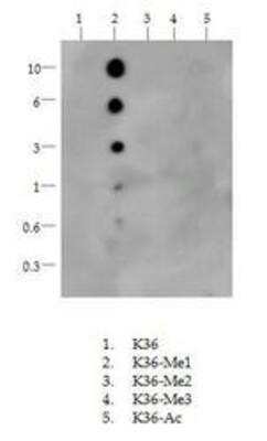
Dot Blot
Immunocytochemistry/ Immunofluorescence
Western Blot
Formulation, Preparation, and Storage
Purification
Formulation
Format
Preservative
Concentration
Shipping
Stability & Storage
Background: Histone H3
Histones are nuclear proteins responsible for the nucleosome structure of the chromosomal fiber in eukaryotes. Changes in chromatin structure play a large role in the regulation of transcription. The chromatin fibers are compacted through the interaction of a linker histone, H1, with the DNA between the nucleosomes to form higher order chromatin structures.
Common histone modifications include methylation of lysine and arginine, acetylation of lysine, phosphorylation of threonine and serine, and sumoylation, biotinylation, and ubiquitylation of lysine. Posttranslational modifications such as acetylation of core histones regulates gene expression, thus altering protein function and regulation (1). Histone H3 is primarily acetylated at lysines 9, 14, 18, and 23 and have a theoretical molecular weight of 15 kDa. Acetylation at lysine 9 and 14 appears to control histone deposition, chromatin assembly and active transcription. Methylation of arginine residues within histone H3 has also been linked to transcription regulation. Histone H3 has been linked to various types of cancer as a biomarker through the aberrant expression of histone deacetylase (HDAC) enzymes and changes to chromatins (2-4).
References
1. Zhang, Y. X., Akumuo, R. C., Espana, R. A., Yan, C. X., Gao, W. J., & Li, Y. C. (2018). The histone demethylase KDM6B in the medial prefrontal cortex epigenetically regulates cocaine reward memory. Neuropharmacology, 141, 113-125. doi:10.1016/j.neuropharm.2018.08.030
2. Nandakumar, V., Hansen, N., Glenn, H. L., Han, J. H., Helland, S., Hernandez, K, ...Meldrum, D. R. (2016). Vorinostat differentially alters 3D nuclear structure of cancer and non-cancerous esophageal cells. Sci Rep, 6, 30593. doi:10.1038/srep30593
3. Zhou, M., Li, Y., Lin, S., Chen, Y., Qian, Y., Zhao, Z., & Fan, H. (2019). H3K9me3, H3K36me3, and H4K20me3 Expression Correlates with Patient Outcome in Esophageal Squamous Cell Carcinoma as Epigenetic Markers. Dig Dis Sci, 64(8), 2147-2157. doi:10.1007/s10620-019-05529-2
4. Li, Y., Guo, D., Sun, R., Chen, P., Qian, Q., & Fan, H. (2019). Methylation Patterns of Lys9 and Lys27 on Histone H3 Correlate with Patient Outcome in Gastric Cancer. Dig Dis Sci, 64(2), 439-446. doi:10.1007/s10620-018-5341-8
Alternate Names
Gene Symbol
UniProt
Additional Histone H3 Products
Product Documents for Histone H3 [Monomethyl Lys36] Antibody - BSA Free
Product Specific Notices for Histone H3 [Monomethyl Lys36] Antibody - BSA Free
Epi-Plus antibody production in collaboration with Rockland Immunochemicals Inc. *Chromatin immunoprecipitation was performed on this product with a standard set of primers that amplify active gene loci (GAPDH, RPL30), inactive loci (MYOD1, AFM), and heterochromatic loci (alphaSat, SAT2). The resulting data indicated there was little to no amplification of these sites, which corresponds with the current literature. Until a more specific locus can be established and validated, we give this product validity for ChIP and it is backed by the full Novus guarantee.
Epi-Plus antibody production in collaboration with Rockland Immunochemicals Inc.
This product is for research use only and is not approved for use in humans or in clinical diagnosis. Primary Antibodies are guaranteed for 1 year from date of receipt.
![Western Blot: Histone H3 [Monomethyl Lys36] Antibody [NB21-1251] Western Blot: Histone H3 [Monomethyl Lys36] Antibody [NB21-1251]](https://resources.bio-techne.com/images/products/Histone-H3-[Monomethyl-Lys36]-Antibody-Western-Blot-NB21-1251-img0010.jpg)
![Immunocytochemistry/ Immunofluorescence: Histone H3 [Monomethyl Lys36] Antibody [NB21-1251] Immunocytochemistry/ Immunofluorescence: Histone H3 [Monomethyl Lys36] Antibody [NB21-1251]](https://resources.bio-techne.com/images/products/Histone-H3-[Monomethyl-Lys36]-Antibody-Immunocytochemistry-Immunofluorescence-NB21-1251-img0013.jpg)
![Western Blot: Histone H3 [Monomethyl Lys36] Antibody [NB21-1251] Western Blot: Histone H3 [Monomethyl Lys36] Antibody [NB21-1251]](https://resources.bio-techne.com/images/products/Histone-H3-[Monomethyl-Lys36]-Antibody-Western-Blot-NB21-1251-img0009.jpg)