Human ALK/CD246 Antibody
R&D Systems, part of Bio-Techne | Catalog # AF4210

Key Product Details
Species Reactivity
Applications
Label
Antibody Source
Product Specifications
Immunogen
Val19-Ser1038
Accession # Q9UM73
Specificity
Clonality
Host
Isotype
Scientific Data Images for Human ALK/CD246 Antibody
Detection of Human ALK/CD246 by Western Blot.
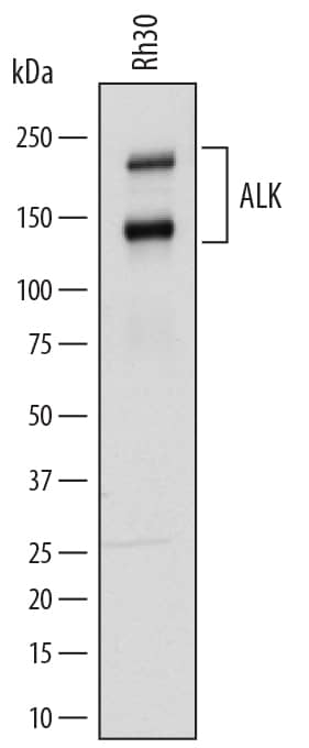
Western blot shows lysates of Rh30 human rhabdomyosarcoma cell line. PVDF membrane was probed with 0.5 µg/mL of Sheep Anti-Human ALK/CD246 Antigen Affinity-purified Polyclonal Antibody (Catalog # AF4210) followed by HRP-conjugated Anti-Sheep IgG Secondary Antibody (HAF016). Specific bands were detected for ALK/CD246 at approximately 220 and 140 kDa (as indicated). This experiment was conducted under reducing conditions and using Immunoblot Buffer Group 1.ALK/CD246 in Human Brain.
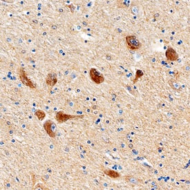
ALK/CD246 was detected in immersion fixed paraffin-embedded sections of human brain (hypothalamus) using Sheep Anti-Human ALK/CD246 Antigen Affinity-purified Polyclonal Antibody (Catalog # AF4210) at 3 µg/mL overnight at 4 °C. Before incubation with the primary antibody, tissue was subjected to heat-induced epitope retrieval using Antigen Retrieval Reagent-Basic (CTS013). Tissue was stained using the Anti-Sheep HRP-DAB Cell & Tissue Staining Kit (brown; CTS019) and counterstained with hematoxylin (blue). Specific staining was localized to neuronal cell bodies. View our protocol for Chromogenic IHC Staining of Paraffin-embedded Tissue Sections.Detection of Human ALK/CD246 by Simple WesternTM.
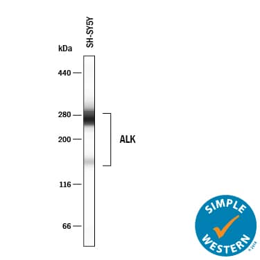
Simple Western lane view shows lysates of SH-SY5Y human neuroblastoma cells, loaded at 0.2 mg/mL. Specific bands were detected for ALK/CD246 at approximately 158 and 266 kDa (as indicated) using 5 µg/mL of Sheep Anti-Human ALK/CD246 Antigen Affinity-purified Polyclonal Antibody (Catalog # AF4210) followed by 1:50 dilution of HRP-conjugated Anti-Sheep IgG Secondary Antibody (Catalog # HAF016). This experiment was conducted under reducing conditions and using the 66-440kDa separation system.Applications for Human ALK/CD246 Antibody
Immunohistochemistry
Sample: Immersion fixed paraffin-embedded sections of human brain (hypothalamus)
Simple Western
Sample: SH-SY5Y human neuroblastoma cells
Western Blot
Sample: Rh30 human rhabdomyosarcoma cell line
Formulation, Preparation, and Storage
Purification
Reconstitution
Formulation
Shipping
Stability & Storage
- 12 months from date of receipt, -20 to -70 °C as supplied.
- 1 month, 2 to 8 °C under sterile conditions after reconstitution.
- 6 months, -20 to -70 °C under sterile conditions after reconstitution.
Background: ALK/CD246
ALK (Anaplastic Lymphoma Kinase; also CD246) is a 200-220 kDa member of the Insulin receptor subfamily, tyrosine kinase family, protein kinase superfamily of proteins. It shows restricted expression, being limited to select sympathetic and sensory neurons, endothelial cells and tumor cells. Although activation of ALK appears to induce cell proliferation, the exact ligand(s) for ALK is unknown. In a temporally-regulated context, midkine has been suggested to bind to ALK, and ionic Zn has been reported to activate ALK cytoplasmically. Mature human ALK is a 1602 amino acid (aa) type I transmembrane glycoprotein (SwissProt #:Q9UM73). It contains a 1020 aa extracellular region (aa 19-1038) that contains one protein-protein MAM domain (aa 264-427), an LDL receptor class A region (aa 437-473), a second MAM domain (aa 480-631) and one utilized phosphorylation site at Ser211. This is accompanied by a long 561 aa cytoplasmic domain that possesses a protein kinase domain (aa 1116-1392) plus eleven utilized Tyr phosphorylation sites. There is one potential splice variant that contains a four aa insert after Glu549. There is also an 80 kDa soluble product that arises from cleavage of the extracellular domain. Although a third 140 kDa product is frequently seen in SDS-Page, its structural basis is unclear. Almost 20 distinct ALK fusion products arise from gene translocations involving multiple intracellular molecules. These seem to involve blocks of aa starting between aa 1050-1060 and continuing to aa 1620. Over aa 19-1038, human ALK shares 88% aa sequence identity with mouse ALK.
Long Name
Alternate Names
Gene Symbol
UniProt
Additional ALK/CD246 Products
Product Documents for Human ALK/CD246 Antibody
Product Specific Notices for Human ALK/CD246 Antibody
For research use only


