Human Angiopoietin-1 Antibody
R&D Systems, part of Bio-Techne | Catalog # MAB923

Key Product Details
Validated by
Species Reactivity
Validated:
Cited:
Applications
Validated:
Cited:
Label
Antibody Source
Product Specifications
Immunogen
Ser20-Phe498
Accession # Q15389
Specificity
Clonality
Host
Isotype
Scientific Data Images for Human Angiopoietin-1 Antibody
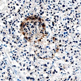
Angiopoietin‑1 in Human Prostate Cancer Tissue.
Angiopoietin-1 was detected in immersion fixed paraffin-embedded sections of human prostate cancer tissue using Mouse Anti-Human Angiopoietin-1 Monoclonal Antibody (Catalog # MAB923) at 15 µg/mL overnight at 4 °C. Tissue was stained using the Anti-Mouse HRP-DAB Cell & Tissue Staining Kit (brown; Catalog # CTS002) and counterstained with hematoxylin (blue). Specific staining was localized to cytoplasm in cancer cells. View our protocol for Chromogenic IHC Staining of Paraffin-embedded Tissue Sections.Detection of Mouse Angiopoietin-1 by Western Blot
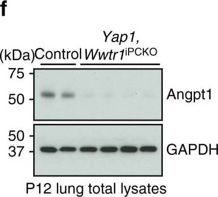
Pericyte-derived Angpt1 controls alveologenesis. a RT-qPCR analysis of Angpt1 and Tie2/Tek expression in freshly sorted lung GFP+, CD31+ or EpCAM+ cells from P7 Pdgfrb(BAC)-CreERT2 R26-mT/mG mice. Data represents mean ± s.e.m. (n = 4 mice). b High magnification images of P10 Angpt1GFP lungs stained for GFP (green), PDGFR beta (red), and PDGFR alpha (blue). Arrows indicate GFP and PDGFR beta double positive pericytes. Scale bar, 15 µm. c RT-qPCR analysis of Angpt1 expression in freshly sorted PDGFR beta+ cells from P7 Yap1,Wwtr1iPCKO and control lungs. Data represents mean ± s.e.m. (n = 4 mice, two-tailed unpaired t-test). dAngpt1 expression in cultured Verteporfin (VP)-treated (48 h) and control pericytes. Data represents mean ± s.e.m. (n = 4, Welch’s t-test). e Expression of the indicated transcripts in freshly sorted CD31+ cells from P7 Yap1,Wwtr1iPCKO and control lungs. Data represents mean ± s.e.m. (n = 4 mice, NS not significant, two-tailed unpaired t-test). f–h Western blot analysis of Angpt1 protein (f; n = 2 controls and 4 mutant mice) and of total and phospho-Tie2 (pTie2) in P12 Yap1,Wwtr1iPCKO and control total lung lysates (g, n = 3 controls and 5 mutants). Molecular weight marker (kDa) is indicated. Relative quantification of signals is shown in h. Two-tailed unpaired t-test. i Scheme showing the time points of tamoxifen administration and analysis for Angpt1iPCKO mice. j, k 3D reconstruction confocal images of P12 Angpt1iPCKO and littermate control lungs stained for AQP5 (green), PDGFR beta (red), and PECAM1 (blue). Panels in k show higher magnification of PECAM1 staining. Scale bar, 50 µm (j) and 30 µm (k). l Quantitation of airspace volume in P12 Angpt1iPCKO and littermate control lung sections with 3D reconstruction surface images. Data represents mean ± s.e.m. (n = 4 mice; p < 0.0001, two-tailed unpaired t-test). m 3D reconstruction confocal images of P12 Angpt1iPCKO and littermate control lungs stained for alphaSMA (red) and tropoelastin (blue). Scale bar, 50 µm. n Quantitation of staining intensity for alphaSMA or tropoelastin shown in m. Intensity was normalized to the size of the parenchymal region. Data represents mean ± s.e.m. (n = 4 mice; NS not significant, two-tailed unpaired t-test). o Schematic summary of findings. Pulmonary pericytes regulate ECs and alveolar epithelial cells via angiocrine factors such as angiopoietin-1 and HGF Image collected and cropped by CiteAb from the following publication (https://pubmed.ncbi.nlm.nih.gov/29934496), licensed under a CC-BY license. Not internally tested by R&D Systems.Detection of Porcine Angiopoietin-1 by Immunohistochemistry
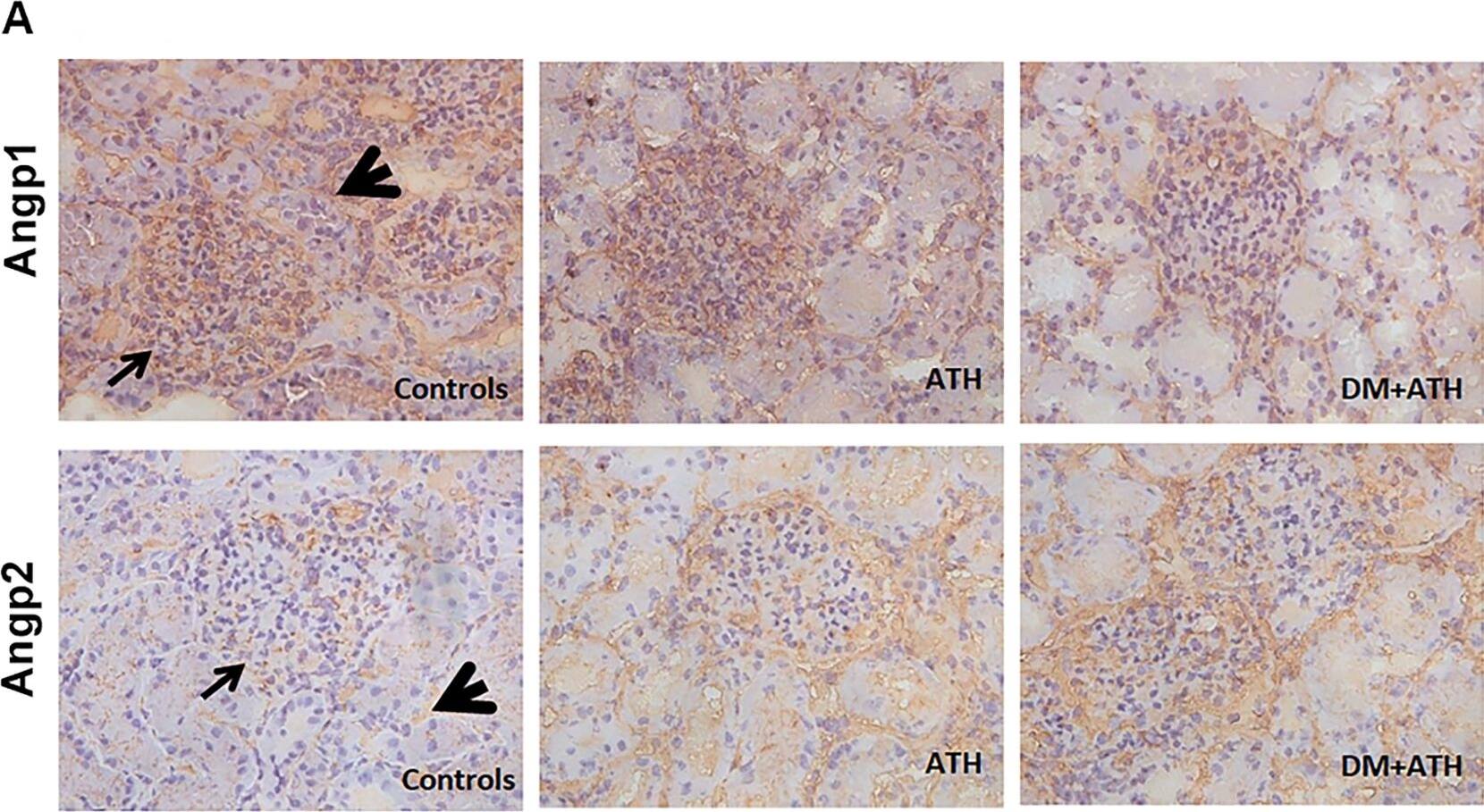
The Angpt2/Angpt1 balance is disturbed in atherogenic DM pigs.A. Representative illustrations of kidney sections stained with Angpt1 (upper panels; arrow: glomerulus; arrowhead: peritubular area) or Angpt2 (lower panels; arrow: glomerulus; arrowhead: tubular staining) in Controls, ATH, and DM+ATH pigs. B. Immunofluorescent double staining of representative kidney sections for desmin (green)/Angpt1 (red;left panel) and vWF (green/Angpt2 (red; middle/right panel). Insets: double positivity for vWF/Angpt2 staining in yellow. C: Quantitative analysis of renal expression of Angpt1, Angpt2 and Angpt2/Angpt1 ratio. D. Relative mRNA expression of Angpt1 and Angpt2. Data are shown as mean ± SEM. *P<0.05 compared to Controls or ATH pigs. Original magnification of A and B: x400. Image collected and cropped by CiteAb from the following publication (https://dx.plos.org/10.1371/journal.pone.0121555), licensed under a CC-BY license. Not internally tested by R&D Systems.Applications for Human Angiopoietin-1 Antibody
Immunohistochemistry
Sample: Immersion fixed paraffin-embedded sections of human prostate cancer tissue
Western Blot
Sample: Recombinant Human Angiopoietin-1 (Catalog # 923-AN)
Reviewed Applications
Read 2 reviews rated 4.5 using MAB923 in the following applications:
Formulation, Preparation, and Storage
Purification
Reconstitution
Formulation
Shipping
Stability & Storage
- 12 months from date of receipt, -20 to -70 °C as supplied.
- 1 month, 2 to 8 °C under sterile conditions after reconstitution.
- 6 months, -20 to -70 °C under sterile conditions after reconstitution.
Background: Angiopoietin-1
Angiopoietin-1 (Ang-1) and Angiopoietin-2 (Ang-2) are two closely related secreted ligands which bind with similar affinity to Tie-2, a receptor tyrosine kinase with immunoglobulin and epidermal growth factor homology domains expressed primarily on endothelial cells and early hematopoietic cells. Tie-2 and angiopoietins have been shown to play critical roles in embryogenic angiogenesis and in maintaining the integrity of the adult vasculature (1). Ang-1 cDNA encodes a 498 amino acid (aa) precursor protein that contains a coiled-coiled domain near the amino-terminus and a fibrinogen-like domain at the C-terminus. Human Ang-1 shares approximately 97% and 60% aa sequence identity with mouse Ang‑1 and human Ang‑2, respectively (1, 2). Ang-1 activates Tie-2 signaling on endothelial cells to promote chemotaxis, cell survival, cell sprouting, vessel growth and stabilization (1, 3, 4). Ang-2 has alternatively been reported to be an antagonist for Ang-1-induced Tie-2 signaling as well as an agonist for Tie-2 signaling, depending on the cell context (5).
References
- Jones, N. et al. (2001) Nat. Rev. Mol. Cell Biol. 2:257.
- Davis, S. et al. (1996) Cell 87:1161.
- Witzenbichler, B. et al. (1998) J. Biol. Chem. 273:18514.
- Papapetropoulos, A. et al. (1999) Lab. Inest. 79:213.
- Teichert-Kuliszewska, K. et al. (2001) Cardiovasc. Res. 49:659.
Alternate Names
Gene Symbol
UniProt
Additional Angiopoietin-1 Products
Product Documents for Human Angiopoietin-1 Antibody
Product Specific Notices for Human Angiopoietin-1 Antibody
For research use only


