Human ATM Antibody
R&D Systems, part of Bio-Techne | Catalog # AF2290

Key Product Details
Validated by
Species Reactivity
Applications
Label
Antibody Source
Product Specifications
Immunogen
Arg2138-Arg2400
Accession # Q13315
Specificity
Clonality
Host
Isotype
Scientific Data Images for Human ATM Antibody
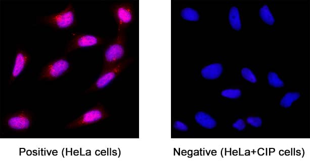
ATM in HeLa Human Cell Line.
ATM was detected in immersion fixed HeLa human cervical epithelial carcinoma cell line (untreated; positive staining) and HeLa human cervical epithelial carcinoma cell line treated with CIP (negative staining) using Sheep Anti-Human ATM Antigen Affinity-purified Polyclonal Antibody (Catalog # AF2290) at 3 µg/mL for 3 hours at room temperature. Cells were stained using the NorthernLights™ 557-conjugated Anti-Rabbit IgG Secondary Antibody (red; NL004) and counterstained with DAPI (blue). Specific staining was localized to cell nuclei. Staining was performed using our protocol for Fluorescent ICC Staining of Non-adherent Cells.ATM in Human Breast.
ATM was detected in immersion fixed paraffin-embedded sections of human breast using Sheep Anti-Human ATM Antigen Affinity-purified Polyclonal Antibody (Catalog # AF2290) at 10 µg/mL overnight at 4 °C. Before incubation with the primary antibody, tissue was subjected to heat-induced epitope retrieval using Antigen Retrieval Reagent-Basic (Catalog # CTS013). Tissue was stained using the Anti-Sheep HRP-DAB Cell & Tissue Staining Kit (brown; Catalog # CTS019) and counterstained with hematoxylin (blue). Specific staining was localized to cytoplasm and nuclei. View our protocol for Chromogenic IHC Staining of Paraffin-embedded Tissue Sections.Applications for Human ATM Antibody
Immunocytochemistry
Sample: Immersion fixed HeLa human cervical epithelial carcinoma cell line
Immunohistochemistry
Sample: Immersion fixed paraffin-embedded sections of human breast
Western Blot
Sample: Recombinant Human ATM.
Formulation, Preparation, and Storage
Purification
Reconstitution
Formulation
Shipping
Stability & Storage
- 12 months from date of receipt, -20 to -70 °C as supplied.
- 1 month, 2 to 8 °C under sterile conditions after reconstitution.
- 6 months, -20 to -70 °C under sterile conditions after reconstitution.
Background: ATM
ATM (Ataxia Telangiectasia Mutated) is a 350-370 kDa member of the ATM subfamily, PI3/PI4-kinase family of enzymes. It is ubiquitously expressed, and serves as a DNA damage sensor. ATM is activated via autophosphorylation at double strand breaks. Following activation, multiple substrates are phosphorylated, including Chk2, and ATR is recruited and activated as part of an integrated repair circuit. Human ATM is 3056 amino acids (aa) in length. It contains one FAT (focal adhesion targeting) domain (aa 1960-2566), a PI-3/PI-4 kinase catalytic domain (aa 2712-2962) and a second, C-terminal FAT domain (aa 3024-3056). There are at least six Ser and four Thr utilized phosphorylation sites, and one critical acetylation activation site at Lys3016. There are at least four potential splice variants. One shows a Trp substitution for aa 536-3056, a second contains an eight aa substitution for aa 2506-3056, a third possesses a five aa substitution for aa 1637-3056, while a fourth contains a premature truncation after Lys2756. Over aa 2138-2400, human ATM shares 82% aa identity with mouse ATM.
Long Name
Alternate Names
Gene Symbol
UniProt
Additional ATM Products
Product Documents for Human ATM Antibody
Product Specific Notices for Human ATM Antibody
For research use only

