Human BCMA/TNFRSF17 Antibody
R&D Systems, part of Bio-Techne | Catalog # MAB107621


Key Product Details
Species Reactivity
Validated:
Cited:
Applications
Validated:
Cited:
Label
Antibody Source
Product Specifications
Immunogen
Met1-Ala54
Accession # Q6PE46
Specificity
Clonality
Host
Isotype
Scientific Data Images for Human BCMA/TNFRSF17 Antibody
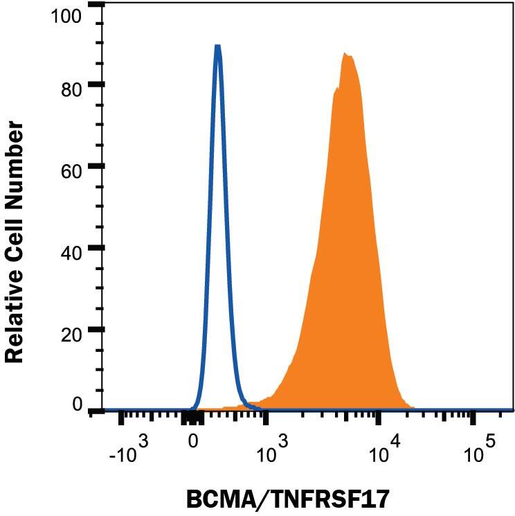
Detection of BCMA/TNFRSF17 in U266 Human Cell Line by Flow Cytometry.
U266 human myeloma cell line was stained with Mouse Anti-Human BCMA/TNFRSF17 Monoclonal Antibody (Catalog # MAB107621, filled histogram) or isotype control antibody (MAB003, open histogram), followed by Phycoerythrin-conjugated Anti-Mouse IgG Secondary Antibody (F0102B). Staining was performed using our Staining Membrane-associated Proteins protocol.Detection of BCMA/TNFRSF17 in RPMI8226 Human Cell Line by Flow Cytometry
RPMI8226 human myeloma cell line was stained with Mouse Anti-Human BCMA/TNFRSF17 Monoclonal Antibody (Catalog # MAB107621, filled histogram) or isotype control antibody (MAB003, open histogram), followed by Phycoerythrin-conjugated Anti-Mouse IgG Secondary Antibody (F0102B). Staining was performed using our Staining Membrane-associated Proteins protocol.BCMA/TNFRSF17 in U266 Human Cell Line .
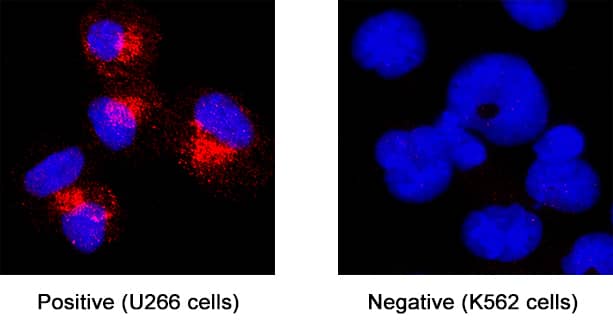
BCMA/TNFRSF17 was detected in immersion fixed U266 human myeloma cell line (positive staining) and K562 human chronic myelogenous leukemia cell line (negative staining) using Mouse Anti-Human BCMA/TNFRSF17 Monoclonal Antibody (Catalog # MAB107621) at 8 µg/mL for 3 hours at room temperature. Cells were stained using the NorthernLights™ 557-conjugated Anti-Mouse IgG Secondary Antibody (red; NL007) and counterstained with DAPI (blue). Specific staining was localized to cytoplasm. Staining was performed using our protocol for Fluorescent ICC Staining of Non-adherent Cells.Applications for Human BCMA/TNFRSF17 Antibody
Flow Cytometry
Sample: RPMI8226 and U266 human myeloma cell lines
Immunocytochemistry
Sample: Immersion fixed U266 human myeloma cell line
Formulation, Preparation, and Storage
Purification
Reconstitution
Formulation
Shipping
Stability & Storage
- 12 months from date of receipt, -20 to -70 °C as supplied.
- 1 month, 2 to 8 °C under sterile conditions after reconstitution.
- 6 months, -20 to -70 °C under sterile conditions after reconstitution.
Background: BCMA/TNFRSF17
BCMA, B cell maturation antigen, is a member of the TNF receptor superfamily. It has been designated TNFRSF17. BCMA is a type III membrane protein containing one extracellular cysteine rich domain. Within the TNFRSF, it shares the highest homology with TACI. BCMA and TACI have both been shown to bind to APRIL and BAFF, members of the TNF ligand superfamily. BCMA expression has been found in immune organs and mature B cell lines. Although some expression has been observed at the cell surface, BCMA appears to be localized to the Golgi compartment. The binding of BCMA to APRIL or BAFF has been shown to stimulate IgM production in peripheral blood B cells and increase the survival of cultured B cells. This data suggests that BCMA may play an important role in B cell development, function and regulation. Human BCMA is a 184 amino acid (aa) protein consisting of a 54 aa extracellular domain, a 23 aa transmembrane domain, and a 107 aa intracellular domain. Mouse and human BCMA share 62% amino acid identity.
References
- Madry, C. et al. (1998) Int. Immunol. 10:1693.
- Gras, M. et al. (1995) Int. Immunol. 7:1093.
- Kwon, B. et al. (1999) Curr. Opin. Immunol. 11:340.
- Marsters, S. et al. (2000) Curr. Biol. 10:785.
- Thompson, J. et al. (2000) J. Exp. Med. 192:129.
Long Name
Alternate Names
Gene Symbol
UniProt
Additional BCMA/TNFRSF17 Products
Product Documents for Human BCMA/TNFRSF17 Antibody
Product Specific Notices for Human BCMA/TNFRSF17 Antibody
For research use only

