Human BDNF Antibody
R&D Systems, part of Bio-Techne | Catalog # MAB248

Key Product Details
Species Reactivity
Validated:
Cited:
Applications
Validated:
Cited:
Label
Antibody Source
Product Specifications
Immunogen
Arg128-Arg247
Accession # P23560
Specificity
Clonality
Host
Isotype
Scientific Data Images for Human BDNF Antibody
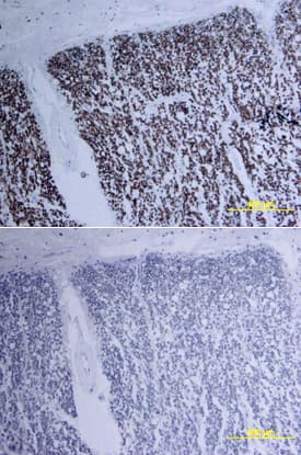
BDNF in Human Spinal Cord.
BDNF was detected in immersion fixed paraffin-embedded sections of human spinal cord using Human BDNF Monoclonal Antibody (Catalog # MAB248) at 15 µg/mL overnight at 4 °C. Tissue was stained using the Anti-Mouse HRP-DAB Cell & Tissue Staining Kit (brown; Catalog # CTS002) and counterstained with hematoxylin (blue). Lower panel shows a lack of labeling if primary antibodies are omitted and tissue is stained only with secondary antibody followed by incubation with detection reagents. View our protocol for Chromogenic IHC Staining of Paraffin-embedded Tissue Sections.Detection of BDNF by Immunohistochemistry
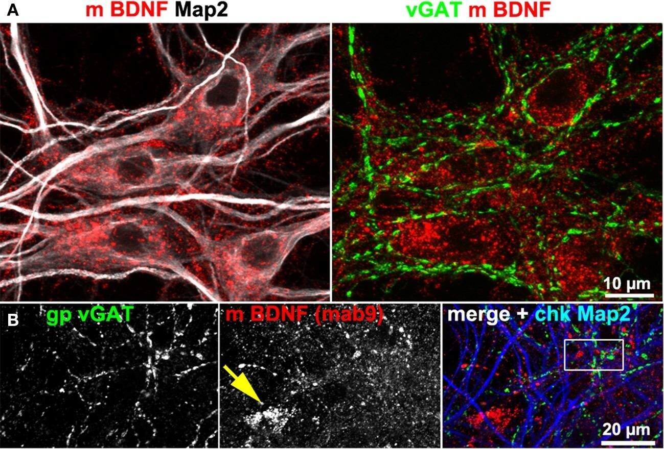
BDNF immunoreactivity is low at GABAergic synapses. (A,B) vGAT and Map2 staining of hippocampal neurons (DIV 25–DIV 31). vGAT+ structures did not show BDNF enrichment. Somatic BDNF staining (yellow arrow in B) was not associated with vGAT+ neurons. (B') (detail in B). Dense anti-BDNF staining (yellow arrowheads) was almost absent from vGAT+ structures (cyan arrowheads). (C–E) In contrast to vGlut-positive structures, vGAT+ neurites barely overlapped with BDNF IR. Cyan arrows point to vGAT+/BDNF-poor synapses. Purple arrowheads point to a close proximity between vGlut and BDNF. Pearson's correlation coefficients describing the overlap between the immune labels are given in the text. Image collected and cropped by CiteAb from the following open publication (https://pubmed.ncbi.nlm.nih.gov/24782711), licensed under a CC-BY license. Not internally tested by R&D Systems.Applications for Human BDNF Antibody
Immunohistochemistry
Sample: Immersion fixed paraffin-embedded sections of human spinal cord
Western Blot
Sample: Recombinant Human BDNF (Catalog # 248-BD)
Formulation, Preparation, and Storage
Purification
Reconstitution
Formulation
Shipping
Stability & Storage
- 12 months from date of receipt, -20 to -70 °C as supplied.
- 1 month, 2 to 8 °C under sterile conditions after reconstitution.
- 6 months, -20 to -70 °C under sterile conditions after reconstitution.
Background: BDNF
Brain-derived neurotrophic factor (BDNF) is a member of the NGF family of neurotrophic factors (also named neurotrophins) that are required for the differentiation and survival of specific neuronal subpopulations in both the central as well as the peripheral nervous system. The neurotrophin family is comprised of at least four proteins including NGF, BDNF, NT-3, and NT-4/5. These secreted cytokines are synthesized as prepropeptides that are proteolytically processed to generate the mature proteins. All neurotrophins have six conserved cysteine residues that are involved in the formation of three disulfide bonds and all share approximately 55% sequence identity at the amino acid level. Similarly to NGF, bioactive BDNF is predicted to be a non-covalently linked homodimer.
BDNF cDNA encodes a 247 amino acid residue precursor protein with a signal peptide and a proprotein that are cleaved to yield the 119 amino acid residue mature BDNF. The amino acid sequence of mature BDNF is identical in all mammals examined. High levels of expression of BDNF have been detected in the hippocampus, cerebellum, fetal eye, and placenta. In addition, low levels of BDNF expression are also found in the pituitary gland, spinal cord, heart, lung, and skeletal muscle. BDNF binds with high affinity and specifically activates the TrkB tyrosine kinase receptor.
References
- Eide, F.F. et al. (1993) Exp. Neurol. 121:200.
- Snider, W.D. (1994) Cell 77:627.
- Barbacid, M. (1994) J. Neurobiol. 25:1386.
Long Name
Alternate Names
Gene Symbol
UniProt
Additional BDNF Products
Product Documents for Human BDNF Antibody
Product Specific Notices for Human BDNF Antibody
For research use only

