Human beta-1,3-Glucuronyltransferase 1/B3GAT1 Antibody
R&D Systems, part of Bio-Techne | Catalog # MAB85603
Human B3GAT1 MAb (Clone 1056734)

Key Product Details
Species Reactivity
Human
Applications
Immunocytochemistry, Immunohistochemistry
Label
Unconjugated
Antibody Source
Monoclonal Mouse IgG2B Clone # 1056734
Product Specifications
Immunogen
Mouse myeloma cell line NS0-derived recombinant rat beta ‑1,3‑Glucuronyltransferase 1/B3GAT1
Asp75-Ile347
Accession # NP_445455
Asp75-Ile347
Accession # NP_445455
Specificity
Detects rat beta ‑1,3‑Glucuronyltransferase 1/B3GAT1 in ELISA
Clonality
Monoclonal
Host
Mouse
Isotype
IgG2B
Scientific Data Images for Human beta-1,3-Glucuronyltransferase 1/B3GAT1 Antibody
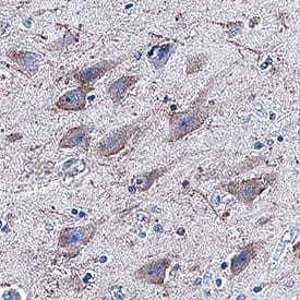
Detection of beta-1,3-Glucuronyltransferase 1/B3GAT1 in Human Brain Hippocampus.
beta-1,3-Glucuronyltransferase 1/B3GAT1 was detected in immersion fixed paraffin-embedded sections of Human Brain Hippocampus using Mouse Anti-Human beta-1,3-Glucuronyltransferase 1/B3GAT1 Monoclonal Antibody (Catalog # MAB85603) at 5 µg/mL for 1 hour at room temperature followed by incubation with the Anti-Mouse IgG VisUCyte™ HRP Polymer Antibody (Catalog # VC001). Before incubation with the primary antibody, tissue was subjected to heat-induced epitope retrieval using VisUCyte Antigen Retrieval Reagent-Basic (Catalog # VCTS021). Tissue was stained using DAB (brown) and counterstained with hematoxylin (blue). Specific staining was localized to cell surface and cytoplasm in neurons. View our protocol for IHC Staining with VisUCyte HRP Polymer Detection Reagents.Detection of beta-1,3-Glucuronyltransferase 1/B3GAT1 in SH-SY5Y (positive) and THP-1 (negative) cells.
beta-1,3-Glucuronyltransferase 1/B3GAT1 was detected in immersion fixed SH-SY5Y human neuroblastoma cell line (positive) and THP-1 human acute monocytic leukemia cell line (negative) cells using Mouse Anti-Human beta-1,3-Glucuronyltransferase 1/B3GAT1 Monoclonal Antibody (Catalog # MAB85603) at 8 µg/mL for 3 hours at room temperature. Cells were stained using the NorthernLights™ 557-conjugated Anti-Mouse IgG Secondary Antibody (red; Catalog # NL007) and counterstained with DAPI (blue). Specific staining was localized to cell surface and cytoplasm. View our protocol for Fluorescent ICC Staining of Non-adherent Cells.Applications for Human beta-1,3-Glucuronyltransferase 1/B3GAT1 Antibody
Application
Recommended Usage
Immunocytochemistry
8-25 µg/mL
Sample: Immersion fixed SH‑SY5Y human neuroblastoma cell line (positive) and THP‑1 human acute monocytic leukemia cell line (negative) cells
Sample: Immersion fixed SH‑SY5Y human neuroblastoma cell line (positive) and THP‑1 human acute monocytic leukemia cell line (negative) cells
Immunohistochemistry
5-25 µg/mL
Sample: Immersion fixed paraffin-embedded sections of Human Brain Hippocampus.
Sample: Immersion fixed paraffin-embedded sections of Human Brain Hippocampus.
Formulation, Preparation, and Storage
Purification
Protein A or G purified from hybridoma culture supernatant
Reconstitution
Reconstitute at 0.5 mg/mL in sterile PBS. For liquid material, refer to CoA for concentration.
Formulation
Lyophilized from a 0.2 μm filtered solution in PBS with Trehalose. *Small pack size (SP) is supplied either lyophilized or as a 0.2 µm filtered solution in PBS.
Shipping
Lyophilized product is shipped at ambient temperature. Liquid small pack size (-SP) is shipped with polar packs. Upon receipt, store immediately at the temperature recommended below.
Stability & Storage
Use a manual defrost freezer and avoid repeated freeze-thaw cycles.
- 12 months from date of receipt, -20 to -70 °C as supplied.
- 1 month, 2 to 8 °C under sterile conditions after reconstitution.
- 6 months, -20 to -70 °C under sterile conditions after reconstitution.
Background: beta-1,3-Glucuronyltransferase 1/B3GAT1
References
- Terayama, K. et al. (1997) Proc. Natl. Acad. Sci. USA 94:6093.
- Shogo, O. et al. (1992) J. Biol. Chem. 267: 22711.
- Kakuda, S. et al. (2005) Glycobiology 2:203.
- Bollensen, E. and Schachner, M. (1987) Neurosci Lett. 82:77.
- Wu, Z.L. et al. (2011) Glycobiology 21:727.
Long Name
UDP-GlcUA:glycoprotein beta-1,3-Glucuronyltransferase
Alternate Names
CD57, GLCATP, GLCUATP, HNK1, LEU7
Gene Symbol
B3GAT1
UniProt
Additional beta-1,3-Glucuronyltransferase 1/B3GAT1 Products
Product Documents for Human beta-1,3-Glucuronyltransferase 1/B3GAT1 Antibody
Product Specific Notices for Human beta-1,3-Glucuronyltransferase 1/B3GAT1 Antibody
For research use only
Loading...
Loading...
Loading...
Loading...

