Human Biglycan Antibody
R&D Systems, part of Bio-Techne | Catalog # AF2667


Key Product Details
Validated by
Species Reactivity
Validated:
Cited:
Applications
Validated:
Cited:
Label
Antibody Source
Product Specifications
Immunogen
Asp38-Lys368
Accession # P21810
Specificity
Clonality
Host
Isotype
Scientific Data Images for Human Biglycan Antibody
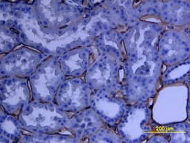
Biglycan in Human Kidney Array.
Biglycan was detected in immersion fixed paraffin-embedded sections of human kidney array using Human Biglycan Antigen Affinity-purified Polyclonal Antibody (Catalog # AF2667) at 15 µg/mL overnight at 4 °C. Tissue was stained using the Anti-Goat HRP-DAB Cell & Tissue Staining Kit (brown; Catalog # CTS008) and counterstained with hematoxylin (blue). View our protocol for Chromogenic IHC Staining of Paraffin-embedded Tissue Sections.Detection of Mouse Biglycan by Immunocytochemistry/Immunofluorescence
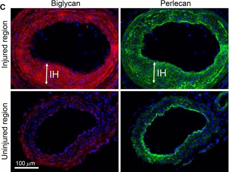
LDL binding to the vessel wall in vitro following treatment with Site B peptide or enzymatic digestion of proteoglycan GAG chains. Tissue sections of carotid arteries from wild‐type mice with intimal hyperplasia were incubated with human LDL. Bound LDL was detected using anti‐apoB antibody. (A) LDL binding to tissue sections pre‐incubated with positively charged SiteB peptide (white circles) or neutrally charged SiteB KE peptide (gray circles). Black circles=no pre‐treatment. White triangles=no LDL incubation (buffer only). (B) LDL binding to tissue sections pre‐treated with the GAG‐degrading enzymes chondroitinase (white circles) or heparinase (white dot circles). Black circles=no pre‐treatment. White triangles=no LDL incubation (buffer only). Data was analyzed using Mann–Whitney rank sum test. Graph bars show median values. P < 0.05 is regarded significant, n = 5–8 in each group. (C) Multi‐immunostaining for biglycan (red, left panels) and perlecan (green, right panels) of tissue sections from the injured region (upper panels) and the uninjured region (lower panels) 3 weeks after carotid injury in a wild‐type mouse. Nuclei are stained with DAPI (blue). Image collected and cropped by CiteAb from the following publication (https://pubmed.ncbi.nlm.nih.gov/28716818), licensed under a CC-BY license. Not internally tested by R&D Systems.Applications for Human Biglycan Antibody
Immunohistochemistry
Sample: Immersion fixed paraffin-embedded sections of human kidney and skin
Western Blot
Sample: Recombinant Human Biglycan (Catalog # 2667-CM)
Formulation, Preparation, and Storage
Purification
Reconstitution
Formulation
Shipping
Stability & Storage
- 12 months from date of receipt, -20 to -70 °C as supplied.
- 1 month, 2 to 8 °C under sterile conditions after reconstitution.
- 6 months, -20 to -70 °C under sterile conditions after reconstitution.
Background: Biglycan
Biglycan is a small secreted proteoglycan belonging to the leucine-rich repeat (LRR) protein family. It contains a central 12 LRR repeat domain flanked by small cysteine clusters at either side. Two glycosaminoglycans (GAG) chains are attached near the amino terminus. Biglycan binds BMP-4 and Chordin to regulate BMP-4 signaling. It may also be involved in collagen fiber assembly. Human Biglycan shares 98% amino acid sequence homology with mouse Biglycan, 97% sequence homology with rat and bovine Biglycan and 96% sequence homology with canine and equine Biglycan.
Alternate Names
Gene Symbol
UniProt
Additional Biglycan Products
Product Documents for Human Biglycan Antibody
Product Specific Notices for Human Biglycan Antibody
For research use only
