Human Calcium Activated Nucleotidase 1/CANT1 Antibody
R&D Systems, part of Bio-Techne | Catalog # AF6720

Key Product Details
Species Reactivity
Applications
Label
Antibody Source
Product Specifications
Immunogen
Gly80-Ile401
Accession # Q8WVQ1
Specificity
Clonality
Host
Isotype
Scientific Data Images for Human Calcium Activated Nucleotidase 1/CANT1 Antibody
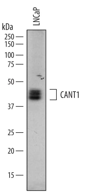
Detection of Human Calcium Activated Nucleotidase 1/CANT1 by Western Blot.
Western blot shows lysates of LNCaP human prostate cancer cell line. PVDF membrane was probed with 0.2 µg/mL of Sheep Anti-Human Calcium Activated Nucleotidase 1/CANT1 Antigen Affinity-purified Polyclonal Antibody (Catalog # AF6720) followed by HRP-conjugated Anti-Sheep IgG Secondary Antibody (Catalog # HAF016). A specific band was detected for Calcium Activated Nucleotidase 1/CANT1 at approximately 40-45 kDa (as indicated). This experiment was conducted under reducing conditions and using Immunoblot Buffer Group 1.Calcium Activated Nucleotidase 1/CANT1 in PC‑3 Human Cell Line.
Calcium Activated Nucleotidase 1/CANT1 was detected in immersion fixed PC-3 human prostate cancer cell line using Sheep Anti-Human Calcium Activated Nucleotidase 1/ CANT1 Antigen Affinity-purified Polyclonal Antibody (Catalog # AF6720) at 10 µg/mL for 3 hours at room temperature. Cells were stained using the NorthernLights™ 637-conjugated Anti-Sheep IgG Secondary Antibody (green; Catalog # NL011) and counterstained with DAPI (blue). Specific staining was localized to perinuclei. View our protocol for Fluorescent ICC Staining of Cells on Coverslips.Applications for Human Calcium Activated Nucleotidase 1/CANT1 Antibody
Immunocytochemistry
Sample: Immersion fixed PC‑3 human prostate cancer cell line
Western Blot
Sample: LNCaP human prostate cancer cell line
Formulation, Preparation, and Storage
Purification
Reconstitution
Formulation
Shipping
Stability & Storage
- 12 months from date of receipt, -20 to -70 °C as supplied.
- 1 month, 2 to 8 °C under sterile conditions after reconstitution.
- 6 months, -20 to -70 °C under sterile conditions after reconstitution.
Background: Calcium Activated Nucleotidase 1/CANT1
CANT1 (soluble Calcium-Activated NucleoTidase 1; also SCAN-1, Apyrase homolog and NF kappaB-activating protein 107) is a 45-48 kDa glycoprotein member of the apyrase (ADP/ATP hydrolysing) family of molecules. It is an ER and Golgi-embedded protein that hydrolyzes UDP in a calcium-dependent manner, and appears to be involved in the regulation of protein glycosylation and folding. It is also reported that CANT1 is essential for cell proliferation. CANT1 is not ubiquitous, but is found in multiple cell types, including prostatic epithelium, fibroblasts, lymphocytes and chondrocytes. Human CANT1 is a 401 amino acid (aa) type II transmembrane glycoprotein. It contains a 44 aa N-terminal cytoplasmic region that possesses an ER retention motif (aa 38-42), and a 339 aa luminal domain (aa 63-401). CANT1 is reported to form membrane-bound disulfide-linked homodimers, and potentially form non-covalent homodimers in a soluble (circulating) state. The soluble form is 35‑40 kDa in size, and presumably cleaved near the transmembrane segment. However, multiple splice forms are possible, and may account for the lower MWs. Two splice forms impact the extracellular region, one which contains a four aa substitution for aa 219-401, and a second that possesses a 26 aa substitution for aa 220‑401. Two additional splice forms utilize alternative start sites at Met 31 and Met165. Over aa 80-401, human CANT1 shares 89% aa sequence identity with mouse CANT1.
Long Name
Alternate Names
Gene Symbol
UniProt
Additional Calcium Activated Nucleotidase 1/CANT1 Products
Product Documents for Human Calcium Activated Nucleotidase 1/CANT1 Antibody
Product Specific Notices for Human Calcium Activated Nucleotidase 1/CANT1 Antibody
For research use only

