Human Catalase Antibody
R&D Systems, part of Bio-Techne | Catalog # MAB3398

Key Product Details
Validated by
Species Reactivity
Validated:
Cited:
Applications
Validated:
Cited:
Label
Antibody Source
Product Specifications
Immunogen
Met1-Leu527
Accession # P04040
Specificity
Clonality
Host
Isotype
Scientific Data Images for Human Catalase Antibody
Detection of Human Catalase by Western Blot.
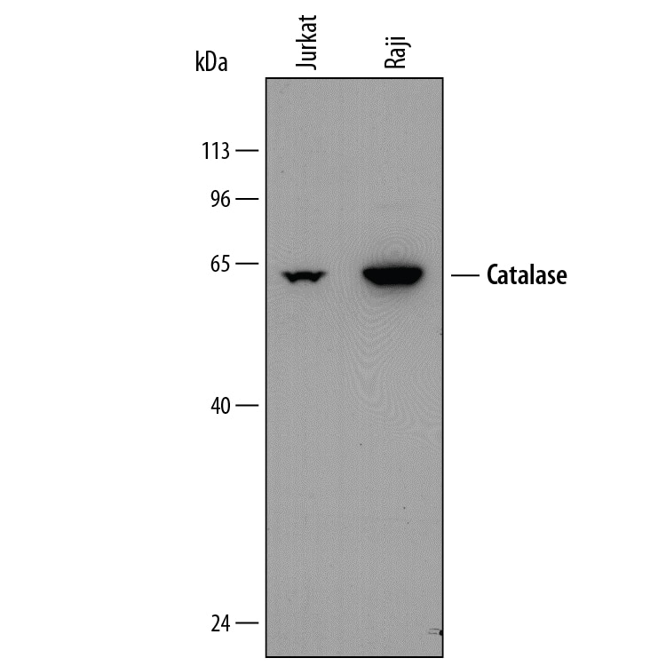
Western blot shows lysates of Jurkat human acute T cell leukemia cell line and Raji human Burkitt's lymphoma cell line. PVDF membrane was probed with 0.5 µg/mL of Mouse Anti-Human Catalase Monoclonal Antibody (Catalog # MAB3398) followed by HRP-conjugated Anti-Mouse IgG Secondary Antibody (Catalog # HAF018). A specific band was detected for Catalase at approximately 64 kDa (as indicated). This experiment was conducted under reducing conditions and using Immunoblot Buffer Group 1.Detection of Human Catalase by Simple WesternTM.
Simple Western lane view shows lysates of Jurkat human acute T cell leukemia cell line, loaded at 0.5 mg/mL. A specific band was detected for Catalase at approximately 61 kDa (as indicated) using 5 µg/mL of Mouse Anti-Human Catalase Monoclonal Antibody (Catalog # MAB3398) . This experiment was conducted under reducing conditions and using the 12-230 kDa separation system.Catalase in HL‑60 Human Cell Line.
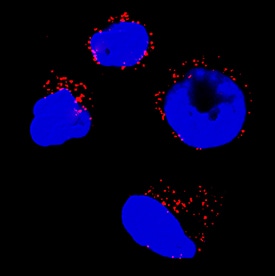
Catalase was detected in immersion fixed HL-60 human acute promyelocytic leukemia cell line using Mouse Anti-Human Catalase Monoclonal Antibody (Catalog # MAB3398) at 3 µg/mL for 3 hours at room temperature. Cells were stained using the NorthernLights™ 557-conjugated Anti-Mouse IgG Secondary Antibody (red; Catalog # NL007) and counterstained with DAPI (blue). Specific staining was localized to peroxisomes. View our protocol for Fluorescent ICC Staining of Non-adherent Cells.Applications for Human Catalase Antibody
Immunocytochemistry
Sample: Immersion fixed HL-60 human acute promyelocytic leukemia cell line
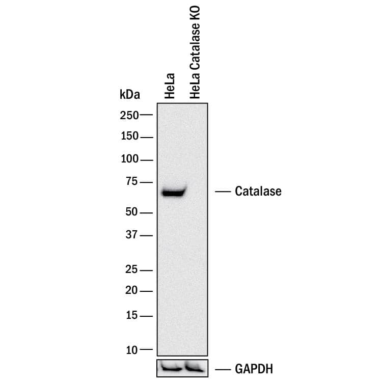
Knockout Validated
Simple Western
Sample: Jurkat human acute T cell leukemia cell line
Western Blot
Sample: Jurkat human acute T cell leukemia cell line and Raji human Burkitt's lymphoma cell line
Formulation, Preparation, and Storage
Purification
Reconstitution
Formulation
Shipping
Stability & Storage
- 12 months from date of receipt, -20 to -70 °C as supplied.
- 1 month, 2 to 8 °C under sterile conditions after reconstitution.
- 6 months, -20 to -70 °C under sterile conditions after reconstitution.
Background: Catalase
Cells have evolved complex mechanisms to maintain redox balance and defend against oxidative stress. Catalase is a tetrameric enzyme comprised of four 60 kDa subunits. Catalase is typically localized in the peroxisome where it functions as an antioxidant, protecting cells from damage due to oxidative stress. Catalase converts reactive oxygen species, such as H2O2, into water and O2. Human Catalase shares 89% homology to mouse and rat Catalase. The cells redox environment can serve as an important signaling switch or trigger to initiate a number of cellular processes, including gene expression, differentiation, proliferation and apoptosis.
Alternate Names
Gene Symbol
UniProt
Additional Catalase Products
Product Documents for Human Catalase Antibody
Product Specific Notices for Human Catalase Antibody
For research use only


