Human CCL19/MIP-3 beta Antibody
R&D Systems, part of Bio-Techne | Catalog # MAB361


Key Product Details
Species Reactivity
Validated:
Cited:
Applications
Validated:
Cited:
Label
Antibody Source
Product Specifications
Immunogen
Gly22-Ser98
Accession # Q99731.1
Specificity
Clonality
Host
Isotype
Scientific Data Images for Human CCL19/MIP-3 beta Antibody
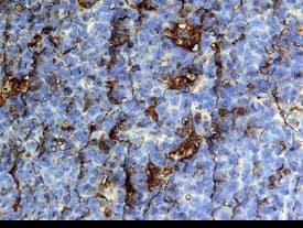
CCL19/MIP-3 beta in Human Tonsil.
CCL19/MIP-3 beta was detected in immersion fixed paraffin-embedded sections of human tonsil using 25 µg/mL Mouse Anti-Human CCL19/ MIP-3 beta Monoclonal Antibody (Catalog # MAB361) overnight at 4 °C. Tissue was stained with the Anti-Mouse HRP-DAB Cell & Tissue Staining Kit (brown; Catalog # CTS002) and counter-stained with hematoxylin (blue). View our protocol for Chromogenic IHC Staining of Paraffin-embedded Tissue Sections.CCL19/MIP-3 beta in Human PBMCs.
CCL19/MIP-3 beta was detected in immersion fixed human peripheral blood mononuclear cells (PBMCs) using 10 µg/mL Mouse Anti-Human CCL19/MIP-3 beta Monoclonal Anti-body (Catalog # MAB361) for 3 hours at room temperature. Cells were stained with the NorthernLights™ 557-conjugated Anti-Mouse IgG Secondary Antibody (red; Catalog # NL007) and counter-stained with DAPI (blue). View our protocol for Fluorescent ICC Staining of Non-adherent Cells.Detection of CCL19/MIP‑3 beta in Human Dendritic Cells by Flow Cytometry.
Human monocyte-derived dendritic cells were stained with Mouse Anti-Human CCL19/MIP-3 beta Monoclonal Antibody (Catalog # MAB361, filled histogram) or isotype control antibody (Catalog # MAB0041, open histogram), followed by Phycoerythrin-conjugated Anti-Mouse IgG Secondary Antibody (Catalog # F0102B). To facilitate intracellular staining, cells were fixed with Flow Cytometry Fixation Buffer (Catalog # FC004) and permeabilized with Flow Cytometry Permeabilization/Wash Buffer I (Catalog # FC005).Applications for Human CCL19/MIP-3 beta Antibody
Immunocytochemistry
Sample: Immersion fixed human peripheral blood mononuclear cells (PBMCs)
Immunohistochemistry
Sample: Immersion fixed paraffin-embedded sections of human tonsil
Intracellular Staining by Flow Cytometry
Sample: Human monocyte-derived dendritic cells
Western Blot
Sample: Recombinant Human CCL19/MIP-3 beta (Catalog # 361-MI)
Reviewed Applications
Read 1 review rated 5 using MAB361 in the following applications:
Formulation, Preparation, and Storage
Purification
Reconstitution
Formulation
Shipping
Stability & Storage
- 12 months from date of receipt, -20 to -70 °C, as supplied.
- 1 month, 2 to 8 °C under sterile conditions after opening.
- 6 months, -20 to -70 °C under sterile conditions after opening.
Background: CCL19/MIP-3 beta
CCL19, also known as MIP-3 beta and ELC (EBI1-Ligand Chemokine), is a 77 amino acid (aa) beta chemokine that is distantly related to other beta chemokines (20-30% aa sequence identity). The gene for MIP-3 beta has been mapped to chromosome 9p13 rather than chromosome 17 where the genes for many human beta chemokines are clustered. MIP-3 beta is constitutively expressed in various lymphoid tissues (including thymus, lymph nodes, appendix and spleen). The expression of MIP-3 beta is
down‑regulated by the anti-inflammatory cytokine IL-10. Recombinant MIP-3 beta is chemotactic for cultured human lymphocytes. MIP-3 beta is a ligand for CCR7 (previously referred to as the Epstein-Barr virus-induced gene 1 (EBI1) orphan receptor), a chemokine receptor that is expressed in various lymphoid tissues and activated B and T lymphocytes. CCR7 is strongly up-regulated in B cells infected with Epstein-Barr virus and T cells infected with herpesvirus 6 or 7.
References
- Yoshida, R. et al. (1997) J. Biol. Chem. 272:13803.
Alternate Names
Gene Symbol
UniProt
Additional CCL19/MIP-3 beta Products
Product Documents for Human CCL19/MIP-3 beta Antibody
Product Specific Notices for Human CCL19/MIP-3 beta Antibody
For research use only

