Human CCR5 Antibody
R&D Systems, part of Bio-Techne | Catalog # MAB181

Key Product Details
Validated by
Biological Validation
Species Reactivity
Validated:
Human
Cited:
Human, Hamster - Cricetulus (Chinese Hamster), Xenograft
Applications
Validated:
Immunohistochemistry
Cited:
Bioassay, Flow Cytometry, Immunocytochemistry, Immunohistochemistry, Immunohistochemistry-Paraffin, Neutralization
Label
Unconjugated
Antibody Source
Monoclonal Mouse IgG2B Clone # 45523
Product Specifications
Immunogen
NS0 mouse myeloma cell line transfected with human CCR5
Met1-Leu352
Accession # P51681
Met1-Leu352
Accession # P51681
Specificity
Can be used to detect CCR5 present on stimulated human PBMCs only after cells are fixed with 2% formaldehyde. It does not detect CCR5 on unfixed human PBMCs. It does not cross-react with CCR1, CCR2, or CCR3. For additional information regarding epitope specificity for this antibody and other R&D Systems anti-human CCR5 antibodies, see Lee, B. et al., 1999, J. Biol. Chem. 274:9617.
Clonality
Monoclonal
Host
Mouse
Isotype
IgG2B
Scientific Data Images for Human CCR5 Antibody
CCR5 in Human Dorsal Root Ganglia.
CCR5 was detected in immersion fixed paraffin-embedded sections of human dorsal root ganglia using 25 µg/mL Human CCR5 Monoclonal Antibody (Catalog # MAB181) overnight at 4 °C. Tissue was stained with the Anti-Mouse HRP-AEC Cell & Tissue Staining Kit (red; Catalog # CTS003) and counterstained with hematoxylin (blue). View our protocol for Chromogenic IHC Staining of Paraffin-embedded Tissue Sections.Detection of Human CCR5 by Immunohistochemistry
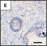
Presence of potential HIV target cells in the human prostate. Immunohistochemistry on uninfected prostate sections before culture showed the presence of periglandular foci of HLA-DR+ (A) and CD4+ cells (B, serial section with A) as well as scattered stromal cells staining positive for HLA-DR (A), CD4 (B), CD3 (C), CD68 (D), CCR5 (E) and CXCR4 (F). The arrows point out immune cells inserted within the epithelium-Scale bars = 50 μm; (G): the respective proportions of CD3, CD4, CD68, CXCR4 and CCR5+ cells per surface unit were evaluated on whole prostate sections from a minimum of 3 donors whose explants were subsequently exposed to HIV-1 strains. Image collected and cropped by CiteAb from the following publication (https://pubmed.ncbi.nlm.nih.gov/19117522), licensed under a CC-BY license. Not internally tested by R&D Systems.Detection of Human CCR5 by Western Blot
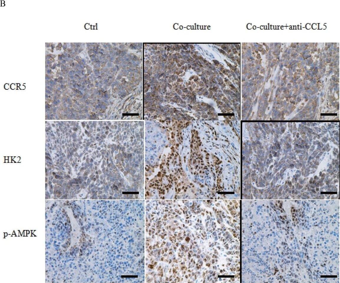
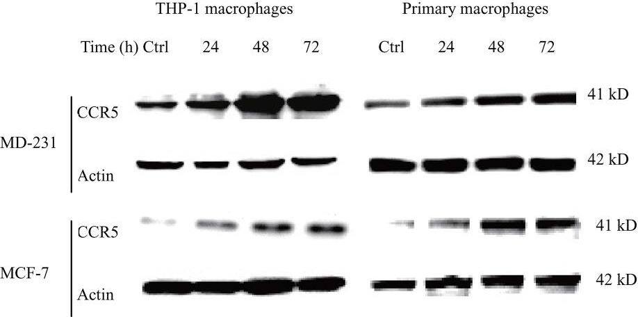
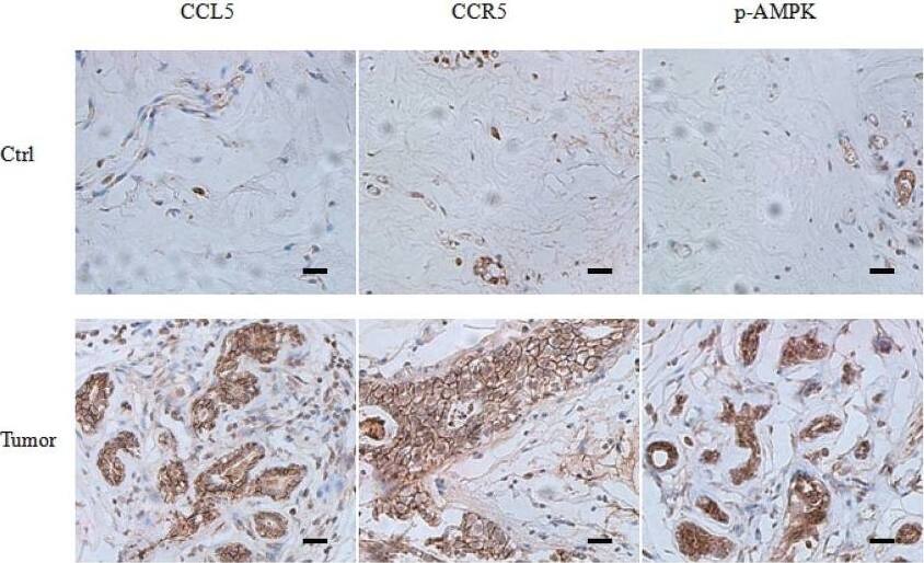
TGF-beta signaling regulated the expression of CCR5(A) 3×10⁵ MDA-MB-231 and MCF-7 cells were stimulated with 1-5ng/ml TGF-beta 1 for 24 h, and total RNA was isolated and tested for CCR5 mRNA by quantitative PCR. (B) Western blot for CCR5 protein in breast cancer cells (106) under TGF-beta 1 stimulation for 48 h. Data presented were representatives of at least three independent experiments. (C) MDA-MB-231 and MCF-7 cells (3×10⁵) were co-transfected with pGL3-CCR5 and pRL-TK and exposed to different concentrations of TGF-beta 1 for 24 h, and luciferase activities were determined. (D) MDA-MB-231 and MCF-7 cells were pre-treated with 5μM SIS3 for 2 h, and cells were subjected to luciferase assay. (E) 106 MCF-7 cells were transfected with TGF betaRI/ALK5 siRNA, and were then co-cultured with lactate-activated THP-1 macrophages (ratio 1:1) for 24 h. The protein levels of CCR5 were assayed by western blot. (F) The expression of TGF-beta 1, CCL5 and CCR5 in clinical samples obtained from breast cancer patients. The mRNA levels were measured by quantitative PCR, and the correlation between TGF-beta 1 and CCL5-CCR5 axis was shown. (G) Representative IHC staining for TGF-beta 1, CCL5 and CCR5 in breast cancer samples. The sample used was derived from 28 breast cancer cases. Scale bars represent 50 μm. *, P<0.05; **, P<0.01. Image collected and cropped by CiteAb from the following publication (https://www.oncotarget.com/lookup/doi/10.18632/oncotarget.22786), licensed under a CC-BY license. Not internally tested by R&D Systems.Applications for Human CCR5 Antibody
Application
Recommended Usage
Immunohistochemistry
8-25 µg/mL
Sample: Immersion fixed paraffin-embedded sections of human dorsal root ganglia
Sample: Immersion fixed paraffin-embedded sections of human dorsal root ganglia
Formulation, Preparation, and Storage
Purification
Protein A or G purified from hybridoma culture supernatant
Reconstitution
Reconstitute at 0.5 mg/mL in sterile PBS. For liquid material, refer to CoA for concentration.
Formulation
Lyophilized from a 0.2 μm filtered solution in PBS with Trehalose. *Small pack size (SP) is supplied either lyophilized or as a 0.2 µm filtered solution in PBS.
Shipping
Lyophilized product is shipped at ambient temperature. Liquid small pack size (-SP) is shipped with polar packs. Upon receipt, store immediately at the temperature recommended below.
Stability & Storage
Use a manual defrost freezer and avoid repeated freeze-thaw cycles.
- 12 months from date of receipt, -20 to -70 °C as supplied.
- 1 month, 2 to 8 °C under sterile conditions after reconstitution.
- 6 months, -20 to -70 °C under sterile conditions after reconstitution.
Background: CCR5
CCR5 is a G protein-linked seven transmembrane domain chemokine receptor. CCR5 serves as a receptor for several chemokines including MIP-1 alpha, MIP-1 beta, MCP-2, and RANTES. It also functions as a coreceptor for Macrophage Tropic HIV-1 infection.
Alternate Names
CCR5, CD195
Gene Symbol
CCR5
UniProt
Additional CCR5 Products
Product Documents for Human CCR5 Antibody
Product Specific Notices for Human CCR5 Antibody
For research use only
Loading...
Loading...
Loading...
Loading...


