Human CD14 Antibody
R&D Systems, part of Bio-Techne | Catalog # AF3831

Key Product Details
Species Reactivity
Applications
Label
Antibody Source
Product Specifications
Immunogen
Thr20-Cys352
Accession # P08571
Specificity
Clonality
Host
Isotype
Scientific Data Images for Human CD14 Antibody
Detection of Human CD14 by Western Blot.
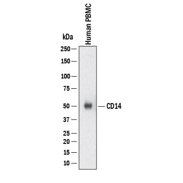
Western blot shows lysates of Human PBMC. PVDF membrane was probed with 0.5 µg/mL of Goat Anti-Human CD14 Antigen Affinity-purified Polyclonal Antibody (Catalog # AF3831) followed by HRP-conjugated Anti-Sheep IgG Secondary Antibody (Catalog # HAF016). A specific band was detected for CD14 at approximately 53 kDa (as indicated). This experiment was conducted under reducing conditions and using Western Blot Buffer Group 1.Detection of Human CD14 by Simple WesternTM.
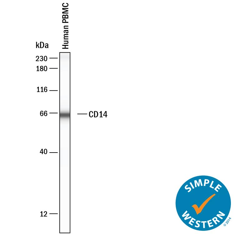
Simple Western lane view shows lysates of Human PBMC, loaded at 0.2 mg/mL. A specific band was detected for CD14 at approximately 65 kDa (as indicated) using 20 µg/mL of Goat Anti-Human CD14 Antigen Affinity-purified Polyclonal Antibody (Catalog # AF3831). This experiment was conducted under reducing conditions and using the 12-230kDa separation system.Detection of CD14 in U937 cells (positive) and MOLT-4 cells (negative).
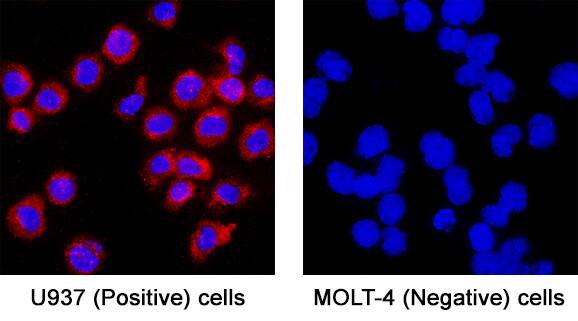
CD14 was detected in immersion fixed U937 human histiocytic lymphoma cells (positive) and absent in MOLT-4 human acute lymphoblastic leukemia cells (negative) using Goat Anti-Human CD14 Antigen Affinity-purified Polyclonal Antibody (Catalog # AF3831) at 5 µg/mL for 3 hours at room temperature. Cells were stained using the NorthernLights™ 557-conjugated Anti-Sheep IgG Secondary Antibody (red; Catalog # NL010) and counterstained with DAPI (blue). Specific staining was localized to cell surface. View our protocol for Fluorescent ICC Staining of Non-adherent Cells.Applications for Human CD14 Antibody
Immunocytochemistry
Sample: Immersion fixed U937 human histiocytic lymphoma cells (positive) and absent in MOLT-4 human acute lymphoblastic leukemia cells (negative)
Simple Western
Sample: Human PBMC
Western Blot
Sample: Human PBMC
Formulation, Preparation, and Storage
Purification
Reconstitution
Formulation
Shipping
Stability & Storage
- 12 months from date of receipt, -20 to -70 °C as supplied.
- 1 month, 2 to 8 °C under sterile conditions after reconstitution.
- 6 months, -20 to -70 °C under sterile conditions after reconstitution.
Background: CD14
CD14 is a 55 kDa cell surface glycoprotein that is preferentially expressed on monocytes/macrophages. The human CD14 cDNA encodes a 375 amino acid (aa) residue precursor protein with a 19 aa signal peptide and a C-terminal hydrophobic region characteristic for glycosylphosphatidyinositol (GPI)-anchored proteins. Human CD14 has four potential N-linked glycosylation sites and also bears O-linked carbohydrates. The amino acid sequence of human CD14 is approximately 65% identical with the mouse, rat, rabbit, and bovine proteins. CD14 is a pattern recognition receptor that binds lipopolysaccharides (LPS) and a variety of ligands derived from different microbial sources. The binding of CD14 with LPS is catalyzed by LPS-binding protein (LBP). The toll-like-receptors have also been implicated in the transduction of CD14-LPS signals. Similar to other GPI-anchored proteins, soluble CD14 can be released from the cell surface by phosphatidyinositol-specific phospholipase C. Soluble CD14 has been detected in serum and body fluids. High concentrations of soluble CD14 have been shown to inhibit LPS-mediated responses. However, soluble CD14 can also potentiate LPS response in cells that do not express cell surface CD14.
References
- Wright, S.D. et al. (1990) Science 249:1431.
- Pugin, J. et al. (1993) Proc. Natl. Acad. Sci. USA 90:2744.
- Beutler, B. (2000) Current Opinion in Immunology 12:20.
- Stelter, F. (2000) Chem. Immunol. 74:25.
Alternate Names
Gene Symbol
UniProt
Additional CD14 Products
Product Documents for Human CD14 Antibody
Product Specific Notices for Human CD14 Antibody
For research use only


