Human CD160 APC-conjugated Antibody
R&D Systems, part of Bio-Techne | Catalog # FAB6700A


Conjugate
Catalog #
Key Product Details
Species Reactivity
Human
Applications
Flow Cytometry
Label
Allophycocyanin (Excitation = 620-650 nm, Emission = 660-670 nm)
Antibody Source
Monoclonal Mouse IgG2B Clone # 688327
Product Specifications
Immunogen
Chinese hamster ovary cell line CHO-derived recombinant human CD160
Ile27-Ser159
Accession # O95971
Ile27-Ser159
Accession # O95971
Specificity
Detects human CD160 in direct ELISAs.
In direct ELISAs and Western blots, no
cross-reactivity with recombinant mouse CD160 is observed.
Clonality
Monoclonal
Host
Mouse
Isotype
IgG2B
Scientific Data Images for Human CD160 APC-conjugated Antibody
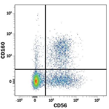
Detection of CD160 in Human PBMCs by Flow Cytometry.
Human peripheral blood mononuclear cells (PBMCs) were stained with Mouse Anti-Human CD160 APC-conjugated Monoclonal Antibody (Catalog # FAB6700A) and Mouse Anti-Human NCAM-1/CD56 PE-conjugated Monoclonal Antibody (Catalog # FAB2408P). View our protocol for Staining Membrane-associated Proteins.Detection of Human CD160 by Flow Cytometry
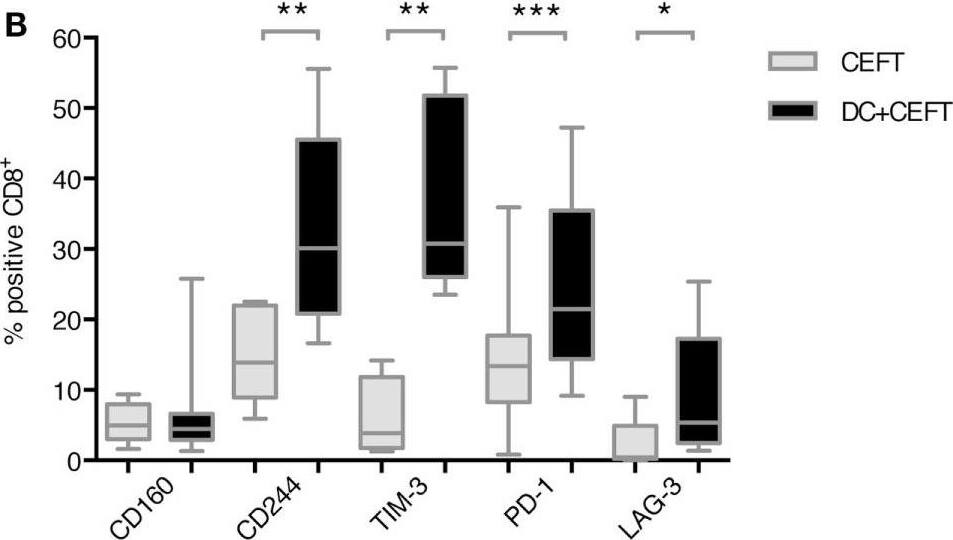
Upregulation of immune checkpoint ligands on T cells after dendritic cell stimulation. T cells of 7–14 healthy donor were cocultured with autologous TLR-3-DCs pulsed with CMV, EBV, influenza, tetanus (CEFT) peptide pool or with CEFT peptide pool alone. Expression of various inhibitory checkpoint molecules was analyzed by flow cytometry. The percentage of positive cells is presented as box-and-whisker plots for CD4+(A) and for CD8+(B) T cells. *p < 0.05; **p < 0.01. Image collected and cropped by CiteAb from the following publication (https://journal.frontiersin.org/article/10.3389/fimmu.2018.00385/full), licensed under a CC-BY license. Not internally tested by R&D Systems.Detection of Human CD160 by Flow Cytometry
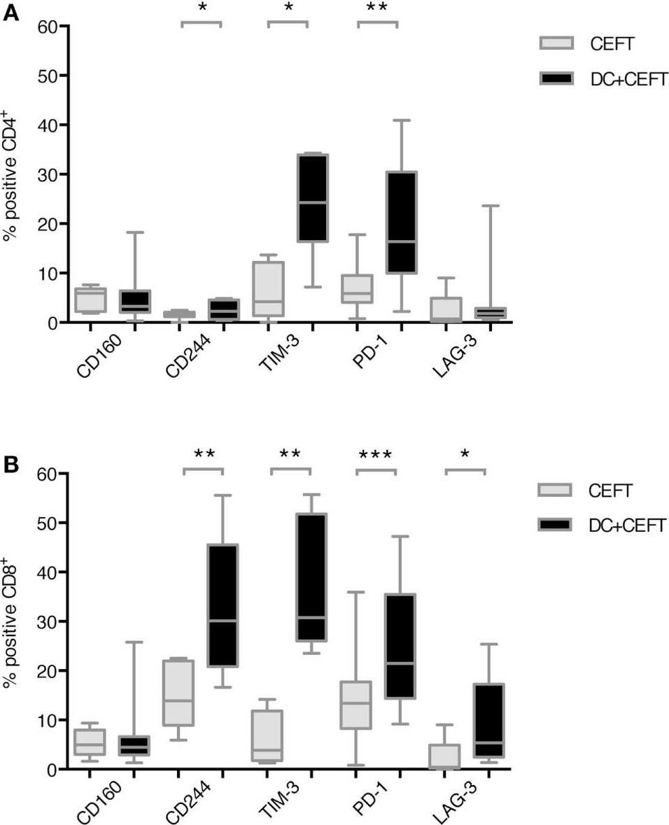
Upregulation of immune checkpoint ligands on T cells after dendritic cell stimulation. T cells of 7–14 healthy donor were cocultured with autologous TLR-3-DCs pulsed with CMV, EBV, influenza, tetanus (CEFT) peptide pool or with CEFT peptide pool alone. Expression of various inhibitory checkpoint molecules was analyzed by flow cytometry. The percentage of positive cells is presented as box-and-whisker plots for CD4+(A) and for CD8+(B) T cells. *p < 0.05; **p < 0.01. Image collected and cropped by CiteAb from the following publication (https://journal.frontiersin.org/article/10.3389/fimmu.2018.00385/full), licensed under a CC-BY license. Not internally tested by R&D Systems.Applications for Human CD160 APC-conjugated Antibody
Application
Recommended Usage
Flow Cytometry
10 µL/106 cells
Sample: Human peripheral blood mononuclear cells (PBMCs)
Sample: Human peripheral blood mononuclear cells (PBMCs)
Formulation, Preparation, and Storage
Purification
Protein A or G purified from hybridoma culture supernatant
Formulation
Supplied in a saline solution containing BSA and Sodium Azide.
Shipping
The product is shipped with polar packs. Upon receipt, store it immediately at the temperature recommended below.
Stability & Storage
Store the unopened product at 2 - 8° C. Do not use past expiration date. Protect from light.
Background: CD160
Alternate Names
BY55, CD160, NK1, NK28
Gene Symbol
CD160
UniProt
Additional CD160 Products
Product Documents for Human CD160 APC-conjugated Antibody
Product Specific Notices for Human CD160 APC-conjugated Antibody
For research use only
Loading...
Loading...
Loading...
Loading...
Loading...

