Human CD28 Antibody
R&D Systems, part of Bio-Techne | Catalog # MAB342


Key Product Details
Species Reactivity
Validated:
Cited:
Applications
Validated:
Cited:
Label
Antibody Source
Product Specifications
Immunogen
Asn19-Pro152
Accession # P10747
Specificity
Clonality
Host
Isotype
Endotoxin Level
Scientific Data Images for Human CD28 Antibody
Human CD28 Antibody Enhances IL-2 Secretion in Jurkat Cells.
Human CD28 Monoclonal Antibody enhances IL-2 secretion in the Jurkat human acute T cell leukemia cell line stimulated with 10 ng/mL phorbol myristate acetate (PMA) and 0.5 µM calcium ionophore, in a dose-dependent manner, as measured using the Quantikine Human IL-2 ELISA Kit (D2050). The ED50 for this effect is typically 0.2-0.6 µg/mL.Detection of CD28 in U266 human myeloma cell line (Positive) & K562 human chronic myelogenous leukemia cell line (Negative).
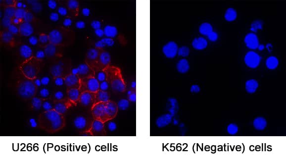
CD28 was detected in immersion fixed U266 human myeloma cell line (Positive) & K562 human chronic myelogenous leukemia cell line (Negative) using Mouse Anti-Human CD28 Monoclonal Antibody (Catalog # MAB342) at 8 µg/mL for 3 hours at room temperature. Cells were stained using the NorthernLights™ 557-conjugated Anti-Mouse IgG Secondary Antibody (red; Catalog # NL007) and counterstained with DAPI (blue). Specific staining was localized to Cytoplasm. View our protocol for Fluorescent ICC Staining of Non-adherent Cells.Applications for Human CD28 Antibody
Agonist Activity
Sample: Jurkat human acute T cell leukemia cell line
Immunocytochemistry
Sample: Immersion fixed U266 human myeloma cell line (Positive) & K562 human chronic myelogenous leukemia cell line (Negative)
Western Blot
Sample: Recombinant Human CD28 Fc Chimera (Catalog # 342-CD)
under non-reducing conditions only
Reviewed Applications
Read 2 reviews rated 5 using MAB342 in the following applications:
Formulation, Preparation, and Storage
Purification
Reconstitution
Formulation
Shipping
Stability & Storage
- 12 months from date of receipt, -20 to -70 °C as supplied.
- 1 month, 2 to 8 °C under sterile conditions after reconstitution.
- 6 months, -20 to -70 °C under sterile conditions after reconstitution.
Background: CD28
CD28 and CTLA-4, together with their ligands, B7-1 and B7-2, constitute one of the dominant costimulatory pathways that regulate T and B cell responses. CD28 and CTLA-4 are structurally homologous molecules that are members of the immunoglobulin (Ig) gene superfamily. Both CD28 and CTLA-4 are composed of a single Ig
V‑like extracellular domain, a transmembrane domain and an intracellular domain. CD28 and CTLA-4 are both expressed on the cell surface as disulfide-linked homodimers or as monomers. The genes encoding these two molecules are closely linked on human chromosome 2 and mouse chromosome 1. Mouse CD28 is expressed constitutively on virtually 100% of mouse T cells and on developing thymocytes. Cell surface expression of mouse CD28 is down-regulated upon ligation of CD28 in the presence of PMA or PHA. In contrast, CTLA-4 is not expressed constitutively but is up-regulated rapidly following T cell activation and CD28 ligation. Cell surface expression of mouse CTLA-4 peaks approximately 48 hours after activation. Although both CTLA-4 and CD28 can bind to the same ligands, CTLA-4 binds to B7-1 and B7-2 with a 20-100 fold higher affinity than CD28. CD28/B7 interaction has been shown to prevent apoptosis of activated T cells via the upregulation of Bcl-xL. CD28 ligation has also been shown to regulate Th1/Th2 differentiation.
References
- Lenschow, D.J. et al. (1996) Annu. Rev. Immunol. 14:233.
- Hathcock, K.S. and R.J. Hodes (1996) Advances in Immunol. 62:131.
- Ward, S.G. (1996) Biochem. J. 318:361.
Alternate Names
Gene Symbol
UniProt
Additional CD28 Products
Product Documents for Human CD28 Antibody
Product Specific Notices for Human CD28 Antibody
For research use only
