Human CD34 Antibody
R&D Systems, part of Bio-Techne | Catalog # AF7227

Key Product Details
Species Reactivity
Validated:
Cited:
Applications
Validated:
Cited:
Label
Antibody Source
Product Specifications
Immunogen
Ser32-Thr290
Accession # P28906
Specificity
Clonality
Host
Isotype
Scientific Data Images for Human CD34 Antibody
Detection of Human CD34 by Western Blot.
Western blot shows lysates of human testies tissue and human thymus tissue. PVDF membrane was probed with 0.5 µg/mL of Sheep Anti-Human CD34 Antigen Affinity-purified Polyclonal Antibody (Catalog # AF7227) followed by HRP-conjugated Anti-Sheep IgG Secondary Antibody (Catalog # HAF016). A specific band was detected for CD34 at approximately 110 kDa (as indicated). This experiment was conducted under reducing conditions and using Immunoblot Buffer Group 1.CD34 in Human Liver.
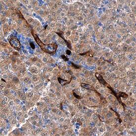
CD34 was detected in immersion fixed paraffin-embedded sections of human liver using Sheep Anti-Human CD34 Antigen Affinity-purified Polyclonal Antibody (Catalog # AF7227) at 1.7 µg/mL overnight at 4 °C. Tissue was stained using the Anti-Sheep HRP-DAB Cell & Tissue Staining Kit (brown; Catalog # CTS019) and counterstained with hematoxylin (blue). Specific staining was localized to endothelial cells in vasculature. View our protocol for Chromogenic IHC Staining of Paraffin-embedded Tissue Sections.Detection of Human CD34 by Immunohistochemistry
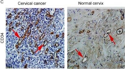
sAng-2 concentration positively relates with Ang-2 expression on the epithelia and MVD in cervical tissues.Representative immunohistochemical staining of Ang-1 (A), Ang-2 (B) and CD34 (C) in 25 cervical cancer tissue specimens and 10 normal controls. Black arrows denote positively stained epithelial cells, whereas red arrows denote positively staining endothelial cells, all appearing brown. Scale bar, 20 µm. (D) sAng-2 is significantly higher in the patients with positive Ang-2 expression on cervix epithelia than those with negative Ang-2 expression. The scatter diagrams show the correlations of sAng-1 (E), sAng-2 (F) and sAng-1/ sAng-2 ratio (G) to MVD in the 35 cervical tissue specimens. Image collected and cropped by CiteAb from the following publication (https://pubmed.ncbi.nlm.nih.gov/28584715), licensed under a CC-BY license. Not internally tested by R&D Systems.Applications for Human CD34 Antibody
Immunohistochemistry
Sample: Immersion fixed paraffin-embedded sections of human liver
Western Blot
Sample: Human testies tissue and Human thymus tissue
Reviewed Applications
Read 1 review rated 5 using AF7227 in the following applications:
Formulation, Preparation, and Storage
Purification
Reconstitution
Formulation
Shipping
Stability & Storage
- 12 months from date of receipt, -20 to -70 °C as supplied.
- 1 month, 2 to 8 °C under sterile conditions after reconstitution.
- 6 months, -20 to -70 °C under sterile conditions after reconstitution.
Background: CD34
CD34 is a 105-115 kDa member of the CD34/podocalyxin family of molecules. It is a sialomucin type glycoprotein, and presents carbohydrate to selectins during cell migration. CD34 is found on mast cells, eosinophils, vascular endothelial cells, stem cells and renal mesangial cells. Mature human CD34 is a 354 amino acid (aa) type I transmembrane protein (aa 32-385). It contains a 259 aa extracellular region (aa 35-287) with utilized N- and O-linked glycosylation sites, and a 74 aa cytoplasmic domain that may undergo Tyr phosphorylation. There is one splice variant that shows a four aa substitution for aa 325-385. Human CD34 can undergo membrane cleavage by bacterial proteases to generate 30-40 kDa soluble fragments. And notably, desialylated CD34 shows a 40 kDa increase in MW (to 150 kDa) when run in SDS-PAGE. Over aa 32-290, human CD34 shares 56% aa identity with mouse CD34.
Alternate Names
Gene Symbol
UniProt
Additional CD34 Products
Product Documents for Human CD34 Antibody
Product Specific Notices for Human CD34 Antibody
For research use only


