Human CD4 Antibody
R&D Systems, part of Bio-Techne | Catalog # MAB11426

Key Product Details
Species Reactivity
Human
Applications
Simple Western, Western Blot
Label
Unconjugated
Antibody Source
Monoclonal Mouse IgG1 Clone # 1068819
Product Specifications
Immunogen
Chinese Hamster Ovary cell line, CHO-derived human CD4
Lys26-Trp390
Accession # P01730
Lys26-Trp390
Accession # P01730
Specificity
Detects human CD4 in direct ELISA.
Clonality
Monoclonal
Host
Mouse
Isotype
IgG1
Scientific Data Images for Human CD4 Antibody
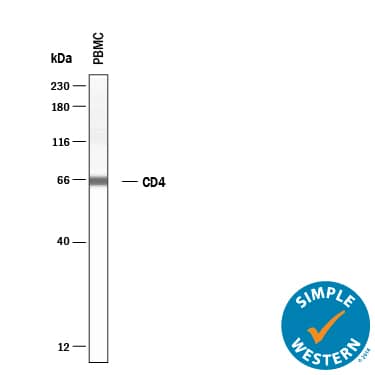
Detection of Human CD4 by Simple WesternTM.
Simple Western lane view shows lysates of Human PBMCs, loaded at 0.2 mg/mL. A specific band was detected for CD4 at approximately 65 kDa (as indicated) using 10 µg/mL of Mouse Anti-Human CD4 Monoclonal Antibody (Catalog # MAB11426) . This experiment was conducted under reducing conditions and using the 12-230 kDa separation system.Detection of Human CD4 by Western Blot.
Western blot shows lysates of PBMC, SUP-T1 human T cell lymphoblastic lymphoma cells. PVDF membrane was probed with 2 µg/mL of Mouse Anti-Human CD4 Monoclonal Antibody (Catalog # MAB11426) followed by HRP-conjugated Anti-Mouse IgG Secondary Antibody (Catalog # HAF018). A specific band was detected for CD4 at approximately 55 kDa (as indicated). This experiment was conducted under reducing conditions and using Western Blot Buffer Group 1.Applications for Human CD4 Antibody
Application
Recommended Usage
Simple Western
10 µg/mL
Sample: Human PBMCs
Sample: Human PBMCs
Western Blot
2 µg/mL
Sample: PBMC, SUP-T1 human T cell lymphoblastic lymphoma cells
Sample: PBMC, SUP-T1 human T cell lymphoblastic lymphoma cells
Formulation, Preparation, and Storage
Purification
Protein A or G purified from hybridoma culture supernatant
Reconstitution
Reconstitute at 0.5 mg/mL in sterile PBS. For liquid material, refer to CoA for concentration.
Formulation
Lyophilized from a 0.2 μm filtered solution in PBS with Trehalose. *Small pack size (SP) is supplied either lyophilized or as a 0.2 µm filtered solution in PBS.
Shipping
Lyophilized product is shipped at ambient temperature. Liquid small pack size (-SP) is shipped with polar packs. Upon receipt, store immediately at the temperature recommended below.
Stability & Storage
Use a manual defrost freezer and avoid repeated freeze-thaw cycles.
- 12 months from date of receipt, -20 to -70 °C as supplied.
- 1 month, 2 to 8 °C under sterile conditions after reconstitution.
- 6 months, -20 to -70 °C under sterile conditions after reconstitution.
Background: CD4
References
- Vignali, D.A.A. (2010) J. Immunol. 184:5933.
- Maddon, P.J. et al. (1985) Cell 42:93.
- Alarcon, B. and H.M. van Santen (2010) Sci. Signal. 3:pe11.
- Wan, Y.Y. and R.A. Flavell (2009) Mol. Cell Biol. 1:20.
- Kitchen, S.G. et al. (2005) Proc. Natl. Acad. Sci. 102:3794.
- Crocker, P.R. et al. (1987) J. Exp. Med. 166:613.
- Biswas, P. et al. (2003) Blood 101:4452.
- Bernstein, H.B. et al. (2006) J. Immunol. 177:3669.
- Funke, I. et al. (1987) J. Exp. Med. 165:1230.
- Doyle, C. and J.L. Strominger (1987) Nature 330:256.
- Huppa, J.B. et al. (2010) Nature 463:963.
- Fragoso, R. et al. (2003) J. Immunol. 170:913.
- Cruikshank, W.W. et al. (1994) Proc. Natl. Acad. Sci. 91:5109.
- Klatzmann, D. et al. (1984) Nature 312:767.
- Dagleish, A.G. et al. (1984) Nature 312:763.
Alternate Names
CD4
Entrez Gene IDs
Gene Symbol
CD4
UniProt
Additional CD4 Products
Product Documents for Human CD4 Antibody
Product Specific Notices for Human CD4 Antibody
For research use only
Loading...
Loading...
Loading...
Loading...
Loading...

