Human CEACAM-16 Antibody
R&D Systems, part of Bio-Techne | Catalog # MAB9885
Recombinant Monoclonal Antibody.


Conjugate
Catalog #
Key Product Details
Species Reactivity
Human
Applications
Flow Cytometry, Immunocytochemistry
Label
Unconjugated
Antibody Source
Recombinant Monoclonal Rabbit IgG Clone # 2747B
Product Specifications
Immunogen
Chinese Hamster Ovary cell line CHO-derived human CEACAM-16
Met1-Gly425
Accession # Q2WEN9
Met1-Gly425
Accession # Q2WEN9
Specificity
Detects human CEACAM-16 in direct ELISAs.
Clonality
Monoclonal
Host
Rabbit
Isotype
IgG
Scientific Data Images for Human CEACAM-16 Antibody
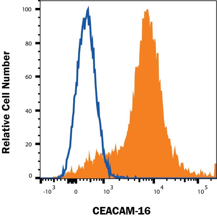
Detection of CEACAM-16 in HEK293 Human Cell Line Transfected with Human CEACAM-16 by Flow Cytometry
HEK293 human embryonic kidney cell line transfected with human CEACAM-16 (filled histogram) or irrelevant protein (open histogram) was stained with Rabbit Anti-Human CEACAM-16 Monoclonal Antibody (Catalog # MAB9885) followed by Allophycocyanin-conjugated Anti-Rabbit IgG Secondary Antibody (F0111). Quadrant markers were set based on control antibody staining (MAB1050). Staining was performed using our Staining Membrane-associated Proteins protocol.CEACAM-16 in HEK293 Human Cell Line.
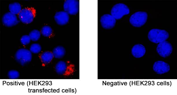
CEACAM-16 was detected in immersion fixed HEK293 human embryonic kidney cell line transfected (positive staining) and HEK293 human embryonic kidney cell line (non-transfected, negative staining) using Rabbit Anti-Human CEACAM-16 Monoclonal Antibody (Catalog # MAB9885) at 3 µg/mL for 3 hours at room temperature. Cells were stained using the NorthernLights™ 557-conjugated Anti-Rabbit IgG Secondary Antibody (red; NL004) and counterstained with DAPI (blue). Specific staining was localized to cytoplasm. Staining was performed using our protocol for Fluorescent ICC Staining of Non-adherent Cells.Applications for Human CEACAM-16 Antibody
Application
Recommended Usage
Flow Cytometry
0.25 µg/106 cells
Sample: HEK293 Human Cell Line Transfected with Human CEACAM-16
Sample: HEK293 Human Cell Line Transfected with Human CEACAM-16
Immunocytochemistry
3-25 µg/mL
Sample: Immersion fixed HEK293 human embryonic kidney cell line transfected
Sample: Immersion fixed HEK293 human embryonic kidney cell line transfected
Formulation, Preparation, and Storage
Purification
Protein A or G purified from cell culture supernatant
Reconstitution
Reconstitute at 0.5 mg/mL in sterile PBS. For liquid material, refer to CoA for concentration.
Formulation
Lyophilized from a 0.2 μm filtered solution in PBS with Trehalose. *Small pack size (SP) is supplied either lyophilized or as a 0.2 µm filtered solution in PBS.
Shipping
Lyophilized product is shipped at ambient temperature. Liquid small pack size (-SP) is shipped with polar packs. Upon receipt, store immediately at the temperature recommended below.
Stability & Storage
Use a manual defrost freezer and avoid repeated freeze-thaw cycles.
- 12 months from date of receipt, -20 to -70 °C as supplied.
- 1 month, 2 to 8 °C under sterile conditions after reconstitution.
- 6 months, -20 to -70 °C under sterile conditions after reconstitution.
Background: CEACAM-16
References
- Beauchemin, N. et al. (1999) Exp. Cell Res. 252:243.
- Zebhauser R. et al. (2005) Genomics 86:566.
- Zheng, J. et al. (2011) PNAS. 108(10):4218.
- Gold P. and Freedman S.O. (1965) J Exp Med 122:467.
- Obrink, B. (1997) Curr Opin Cell Biol 9:616.
- Horst, A.K. and Wagener, C. (2004) Handb Exp Pharmacol 283.
- Kuespert K et al. (2006) Curr Opin Cell Biol. 18(5):565.
- Wang, H. et al. (2015) J Hum Genet. 60(3):119.
Long Name
Carcinoembryonic Antigen-related Cell Adhesion Molecule 16
Alternate Names
CEAL2, DFNA4B
Gene Symbol
CEACAM16
UniProt
Additional CEACAM-16 Products
Product Documents for Human CEACAM-16 Antibody
Product Specific Notices for Human CEACAM-16 Antibody
For research use only
Loading...
Loading...
Loading...
Loading...
Loading...
