Human CLEC9a Antibody
R&D Systems, part of Bio-Techne | Catalog # AF6049

Key Product Details
Species Reactivity
Validated:
Cited:
Applications
Validated:
Cited:
Label
Antibody Source
Product Specifications
Immunogen
Lys57-Val241
Accession # Q6UXN8
Specificity
Clonality
Host
Isotype
Endotoxin Level
Scientific Data Images for Human CLEC9a Antibody
Detection of Human CLEC9a by Western Blot.
Western blot shows lysates of human CD14+peripheral blood mononuclear cells (PBMC). PVDF Membrane was probed with 1 µg/mL of Sheep Anti-Human CLEC9a Antigen Affinity-purified Polyclonal Antibody (Catalog # AF6049) followed by HRP-conjugated Anti-Sheep IgG Secondary Antibody (Catalog # HAF016). A specific band was detected for CLEC9a at approximately 45 kDa (as indicated). This experiment was conducted under reducing conditions and using Immunoblot Buffer Group 8.CLEC9a in Human Spleen.
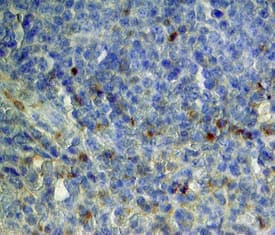
CLEC9a was detected in immersion fixed paraffin-embedded sections of human spleen using Sheep Anti-Human CLEC9a Antigen Affinity-purified Polyclonal Antibody (Catalog # AF6049) at 15 µg/mL overnight at 4 °C. Tissue was stained using the Anti-Sheep HRP-DAB Cell & Tissue Staining Kit (brown; Catalog # CTS019) and counterstained with hematoxylin (blue). Specific staining was localized to cytoplasm. View our protocol for Chromogenic IHC Staining of Paraffin-embedded Tissue Sections.Applications for Human CLEC9a Antibody
Immunohistochemistry
Sample: Immersion fixed paraffin-embedded sections of human spleen
Western Blot
Sample: Human CD14+ peripheral blood mononuclear cells (PBMC).
Binding Inhibition
Formulation, Preparation, and Storage
Purification
Reconstitution
Formulation
Shipping
Stability & Storage
- 12 months from date of receipt, -20 to -70 °C as supplied.
- 1 month, 2 to 8 °C under sterile conditions after reconstitution.
- 6 months, -20 to -70 °C under sterile conditions after reconstitution.
Background: CLEC9a
CLEC9a (C-type lectin domain family 9 member A), also known as DNGR-1, is a type II transmembrane glycoprotein member of the C-type lectin superfamily (1, 2). Mature human CLEC9a consists of a 35 amino acid (aa) cytoplasmic domain with an ITAM-like motif, a 21 aa transmembrane segment, and a 185 extracellular domain (ECD) that contains a stalk region and one C-type lectin domain (CTLD) (3‑5). Within the ECD, human CLEC9a shares 57% aa sequence identity with mouse and rat CLEC9a. Although the CTLD of CLEC9a structurally resembles that of other C-type lectins, it lacks the conserved residues that typically mediate calcium and carbohydrate binding. CLEC9a is expressed as a disulfide-linked homodimer of approximately 45-50 kDa N-glycosylated subunits (3, 5). Human CLEC9a expression is restricted to a subpopulation of BDCA-3+ conventional dendritic cells (cDC) and CD16- monocytes (3‑5). BDCA-3+ cDC are analagous to mouse CD8+ cDC which are specialized in antigenic cross-presentation in complex with MHC class I molecules (6). In mouse, CLEC9a is additionally expressed on plasmacytoid dendritic cells (4, 5). CLEC9a ligation enhances antigen uptake and processing, leading to presentation on MHC class I and cytotoxic T cell (CTL) priming (3‑5). In mouse, CLEC9a recognizes normally intracellular determinant(s) of necrotic cells and mediates their uptake by the dendritic cell (7). The subsequent antigenic cross-presentation to CTL is important for clearing necrotic cellular debris (7). CLEC9a signaling triggers activation of the tyrosine kinase Syk (3, 7).
References
- Huysamen, C. and G.D. Brown (2009) FEMS Microbiol. Lett. 290:121.
- Geijtenbeek, T.B.H. et al. (2004) Annu. Rev. Immunol. 22:33.
- Huysamen, C. et al. (2008) J. Biol. Chem. 283:16693.
- Caminschi, I. et al. (2008) Blood 112:3264.
- Sancho, D. et al. (2008) J. Clin. Invest. 118:2098.
- Dudziak, D. et al. (2007) Science 315:107.
- Sancho, D. et al. (2009) Nature 458:899.
Long Name
Alternate Names
Gene Symbol
UniProt
Additional CLEC9a Products
Product Documents for Human CLEC9a Antibody
Product Specific Notices for Human CLEC9a Antibody
For research use only

