Human CXCL9/MIG Antibody
R&D Systems, part of Bio-Techne | Catalog # AF392


Key Product Details
Species Reactivity
Validated:
Cited:
Applications
Validated:
Cited:
Label
Antibody Source
Product Specifications
Immunogen
Thr23-Thr125
Accession # Q07325
Specificity
Clonality
Host
Isotype
Endotoxin Level
Scientific Data Images for Human CXCL9/MIG Antibody
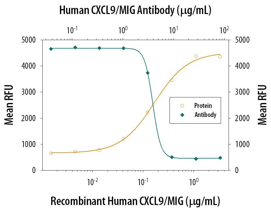
Chemotaxis Induced by CXCL9/MIG and Neutral-ization by Human CXCL9/ MIG Antibody.
Recombinant Human CXCL9/ MIG (392-MG) chemo-attracts the BaF3 mouse pro-B cell line transfected with mouse CXCR3 in a dose-dependent manner (orange line). The amount of cells that migrated through to the lower chemotaxis chamber was measured by Resazurin (AR002). Chemotaxis elicited by Recombinant Human CXCL9/ MIG (1 µg/mL) is neutralized (green line) by increasing concentrations of Goat Anti-Human CXCL9/MIG Antigen Affinity-purified Polyclonal Antibody (Catalog # AF392). The ND50 is typically 5-40 µg/mL.CXCL9/MIG in THP‑1 Human Cell Line.
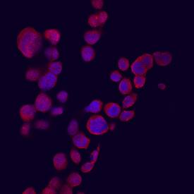
CXCL9/MIG was detected in immersion fixed THP-1 human acute monocytic leukemia cell line stimulated with IFN-gamma using Goat Anti-Human CXCL9/MIG Antigen Affinity-purified Polyclonal Antibody (Catalog # AF392) at 10 µg/mL for 3 hours at room temperature. Cells were stained using the Northern-Lights™ 557-conjugated Anti-Goat IgG Secondary Antibody (red; Catalog # NL001) and counter-stained with DAPI (blue). View our protocol for Fluorescent ICC Staining of Cells on Coverslips.Detection of Human CXCL9/MIG by Immunohistochemistry
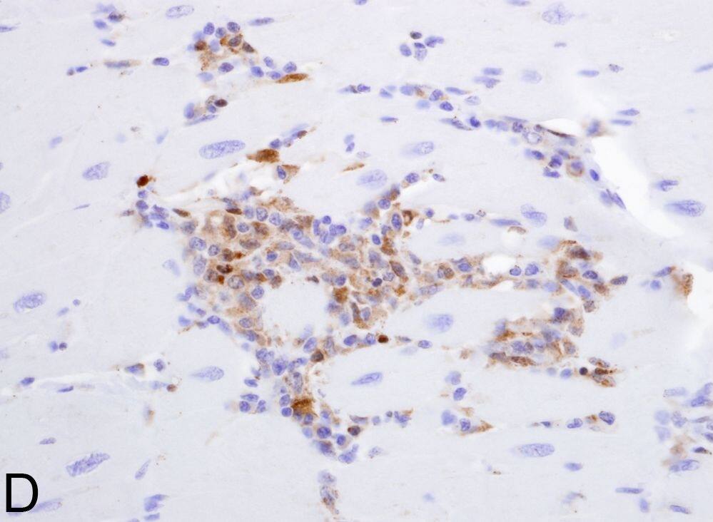
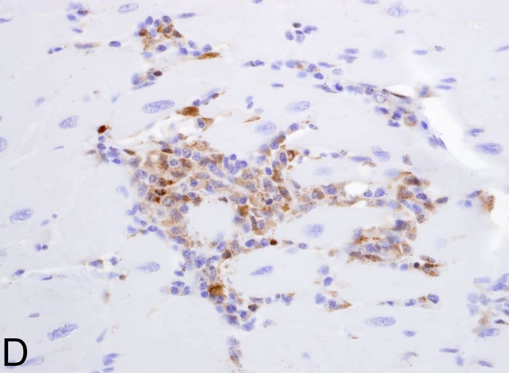
CD8+ T- and CD20+ B-lymphocytes are not localized within inflammatory foci.Immunohistochemistry using anti-CD20 identified a few B-lymphocytes in the inflammatory foci (arrows) (A). In contrast, no CD8+ T-lymphocytes are localized using anti-CD8 antibody (B) (400X). Immunohistrochemistry using anti-pro-IL-18 (C) or anti-CXCL9 (D) reveals the expression of these molecules on mononuclear cells within inflammatory foci (200X). Image collected and cropped by CiteAb from the following open publication (https://dx.plos.org/10.1371/journal.pone.0014429), licensed under a CC-BY license. Not internally tested by R&D Systems.Applications for Human CXCL9/MIG Antibody
Immunocytochemistry
Sample: Immersion fixed human peripheral blood mononuclear cells treated with PMA and calcium ionomycin, and THP-1 human acute monocytic leukemia cell line stimulated with IFN-gamma
Western Blot
Sample: Recombinant Human CXCL9/MIG (Catalog # 392-MG)
Neutralization
Formulation, Preparation, and Storage
Purification
Reconstitution
Formulation
Shipping
Stability & Storage
- 12 months from date of receipt, -20 to -70 °C as supplied.
- 1 month, 2 to 8 °C under sterile conditions after reconstitution.
- 6 months, -20 to -70 °C under sterile conditions after reconstitution.
Background: CXCL9/MIG
CXCL9, a member of the alpha subfamily of chemokines that lack the ELR domain, was initially identified as a lymphokine-activated gene in mouse macrophages. Human CXCL9 was subsequently cloned using mouse MIG cDNA as a probe. The CXCL9 gene is induced in macrophages and in primary glial cells of the central nervous system specifically in response to IFN-gamma. CXCL9 has been shown to be a chemoattractant for activated T-lymphocytes and TIL but not for neutrophils or monocytes. The human CXCL9 cDNA encodes a 125 amino acid residue precursor protein with a 22 amino acid residue signal peptide that is cleaved to yield a 103 amino acid residue mature protein. CXCL9 has an extended carboxy-terminus containing greater than 50% basic amino acid residues and is larger than most other chemokines. The carboxy-terminal residues of CXCL9 are prone to proteolytic cleavage resulting in size heterogeneity of natural and recombinant CXCL9. CXCL9 with large carboxy-terminal deletions have been shown to have diminished activity in the calcium flux assay. A chemokine receptor (CXCR3) specific for CXCL9 and IP-10 has been cloned and shown to be highly expressed in IL-2-activated T-lymphocytes. The E. coli-expressed CXCL9 preparations produced at R&D Systems have been shown to contain greater than 80% full length CXCL9.
References
- Loetscher, M. et al. (1996) J. Exp. Med. 184:963.
- Liao, F. et al. (1995) J. Exp. Med. 182:1301.
- Vanguri, P. (1995) J. Neuroimmunol. 56:35.
Alternate Names
Gene Symbol
UniProt
Additional CXCL9/MIG Products
Product Documents for Human CXCL9/MIG Antibody
Product Specific Notices for Human CXCL9/MIG Antibody
For research use only


