Human CXCR4 Antibody
R&D Systems, part of Bio-Techne | Catalog # MAB173


Conjugate
Catalog #
Key Product Details
Species Reactivity
Validated:
Human
Cited:
Human, Mouse, Feline
Applications
Validated:
CyTOF-ready, Flow Cytometry, Neutralization
Cited:
Binding Assay, Blocking, Flow Cytometry, Immunocytochemistry, Immunohistochemistry, Immunohistochemistry-Paraffin, Neutralization, Surface Plasmon Resonance, Western Blot
Label
Unconjugated
Antibody Source
Monoclonal Mouse IgG2B Clone # 44717
Product Specifications
Immunogen
3T3 cells transfected with human CXCR4
Specificity
Reacts specifically with human and non-human cells expressing human CXCR4 (fusin) as detected by flow cytometry. It will also react with cells expressing feline CXCR4 but not rat CXCR4. This antibody does not cross-react with other chemokine receptors.
Clonality
Monoclonal
Host
Mouse
Isotype
IgG2B
Endotoxin Level
<0.10 EU per 1 μg of the antibody by the LAL method.
Scientific Data Images for Human CXCR4 Antibody
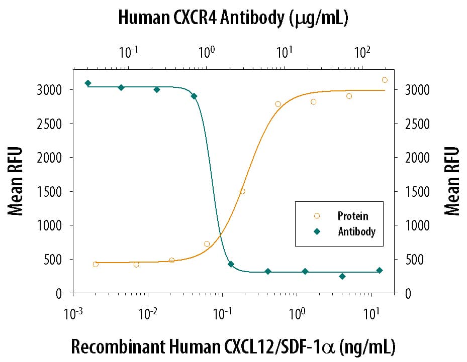
Chemotaxis Induced by CXCL12/SDF‑1 alpha and Neutralization by Human CXCR4 Antibody.
Recombinant Human/Feline/Rhesus Macaque CXCL12/SDF-1a (Catalog # 350-NS) chemoattracts the BaF3 mouse pro-B cell line transfected with human CXCR4 in a dose-dependent manner (orange line). The amount of cells that migrated through to the lower chemotaxis chamber was measured by Resazurin (Catalog # AR002). Chemotaxis elicited by Recombinant Human/Feline/Rhesus Macaque CXCL12/SDF-1a (1 ng/mL) is neutralized (green line) by increasing concentrations of Mouse Anti-Human CXCR4 Monoclonal Antibody (Catalog # MAB173). The ND50 is typically 1-5 µg/mL.Detection of Canine CXCR4 by Immunocytochemistry/Immunofluorescence
Ectopic expression of CXCR7 and CXCR4 on MDCK cells.(A) MDCK were stably transfected with empty vector (Mock), with CXCR7, CXCR4, a vector coding for a CXCR7 lacking the cytoplasmic C-terminus ( deltaCXCR7), and a vector coding for a chimeric CXCR7 containing the DRYLAIV motive of CXCR4. Receptor expression was determined by FACS analysis using saturating antibody concentrations (see Methods). (B) Confocal immunofluorescence analysis of unfixed MDCK cells expressing CXCR7 (upper panels) or CXCR4 (lower panels). Cells also expressed N-ter-Lck mCherry as membrane marker (red fluorescence). Left panels: confocal images of planes cut through intracellular regions of MDCK monolayers. Right panels: x-z planes reconstructed from confocal x-y stacks. For receptor (green) detection anti-CXCR7 (11G8 R&D) or anti CXCR4 (MAB173 R&D) were used. Receptor-bound primary antibodies were revealed with goat anti mouse IgG conjugated with Alexa488 (green fluorescence). Image collected and cropped by CiteAb from the following publication (https://pubmed.ncbi.nlm.nih.gov/20161793), licensed under a CC-BY license. Not internally tested by R&D Systems.Detection of CXCR4 by Western Blot
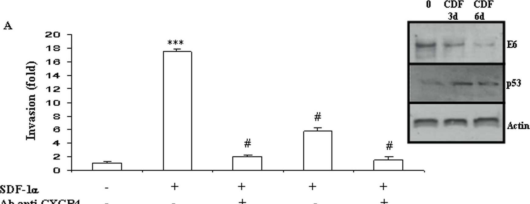
In vitro cell invasion is stimulated by the SDF-1/CXCR4 pathway independently from HPV status.The modulation of E6 expression was monitored using Western-blot in HPV-positive HeLa (A) and TC-1 (B) cells and in HPV-negative B16F10 (C) cells, after 3 and 6 days of incubation with Cidofovir (CDF). Modulation of P53 expression was assessed in HeLa cells after CDF incubation (A). Cell invasion was measured using a Matrigel assay in HeLa (A), TC-1 (B) and B16F10 (C) cells. Recombinant human CXCL12/SDF-1 (100 ng/mL; R&D Systems) was used as chemoattractant and modulation of cell migration was recorded after treatment with CXCR4-blocking antibody or/and Cidofovir (CDF). The invasion rate was determined by counting crystal violet-stained cells. Invasion was stimulated by SDF-1 alpha/CXCR4 independently from the HPV status of the cells but Cidofovir anti-invasive action was restricted to the two HPV-positive cell lines. Three independent experiments with three chambers each time were performed. ***P<0.01, for a statistically true difference, as compared to the untreated group. # P<0.01 compared to SDF-1 alpha-treated group. Image collected and cropped by CiteAb from the following open publication (https://pubmed.ncbi.nlm.nih.gov/19325708), licensed under a CC-BY license. Not internally tested by R&D Systems.Applications for Human CXCR4 Antibody
Application
Recommended Usage
CyTOF-ready
Ready to be labeled using established conjugation methods. No BSA or other carrier proteins that could interfere with conjugation.
Flow Cytometry
2.5 µg/106 cells
Sample: Jurkat human acute T cell leukemia cell line
Sample: Jurkat human acute T cell leukemia cell line
Neutralization
Measured by its ability to neutralize CXCL12/SDF‑1 alpha-induced chemotaxis in the BaF3 mouse pro‑B cell line transfected with human CXCR4. The Neutralization Dose (ND50) is typically 1-5 µg/mL in the presence of 1 ng/mL Recombinant Human/Feline/Rhesus Macaque CXCL12/SDF‑1 alpha.
Formulation, Preparation, and Storage
Purification
Protein A or G purified from hybridoma culture supernatant
Reconstitution
Reconstitute at 0.5 mg/mL in sterile PBS. For liquid material, refer to CoA for concentration.
Formulation
Lyophilized from a 0.2 μm filtered solution in PBS with Trehalose. *Small pack size (SP) is supplied either lyophilized or as a 0.2 µm filtered solution in PBS.
Shipping
Lyophilized product is shipped at ambient temperature. Liquid small pack size (-SP) is shipped with polar packs. Upon receipt, store immediately at the temperature recommended below.
Stability & Storage
Use a manual defrost freezer and avoid repeated freeze-thaw cycles.
- 12 months from date of receipt, -20 to -70 °C as supplied.
- 1 month, 2 to 8 °C under sterile conditions after reconstitution.
- 6 months, -20 to -70 °C under sterile conditions after reconstitution.
Background: CXCR4
References
- Orsini, M.J. et al. (1999) J. Biol. Chem. 274:31076.
- Zagzag, D. et al. (2005) Cancer Res. 65:6178.
- Speetjens, F.M. et al. (2009) Cancer Microenvironment 2:1.
- Wang, L. et al. (2009) Oncology Reports 22:1333.
- Amara, S. et al. (2015) Cancer Biomark. 15:869.
Long Name
C-X-C Motif Chemokine Receptor 4
Alternate Names
CD184, D2S201E, FB22, Fusin, HM89, HSY3RR, LAP-3, LAP3, LCR1, LESTR, NPY3R, NPYRL, NPYY3R, WHIMS
Gene Symbol
CXCR4
Additional CXCR4 Products
Product Documents for Human CXCR4 Antibody
Product Specific Notices for Human CXCR4 Antibody
For research use only
Loading...
Loading...
Loading...
Loading...
Loading...
Loading...


