Human Cystatin B Antibody
R&D Systems, part of Bio-Techne | Catalog # MAB1408

Key Product Details
Validated by
Species Reactivity
Validated:
Cited:
Applications
Validated:
Cited:
Label
Antibody Source
Product Specifications
Immunogen
aa Met2-Phe98
Accession # P04080
Specificity
Clonality
Host
Isotype
Scientific Data Images for Human Cystatin B Antibody
Detection of Human Cystatin B by Western Blot.
Western blot shows lysates of human colon tissue, SK-BR-3 human breast cancer cell line, and A549 human lung carcinoma cell line. PVDF membrane was probed with 2 µg/mL of Mouse Anti-Human Cystatin B Monoclonal Antibody (Catalog # MAB1408) followed by HRP-conjugated Anti-Mouse IgG Secondary Antibody (Catalog # HAF018). A specific band was detected for Cystatin B at approximately 12 kDa (as indicated). This experiment was conducted under reducing conditions and using Immunoblot Buffer Group 1.Neutralization of Cystatin B Activity by Human Cystatin B Antibody.
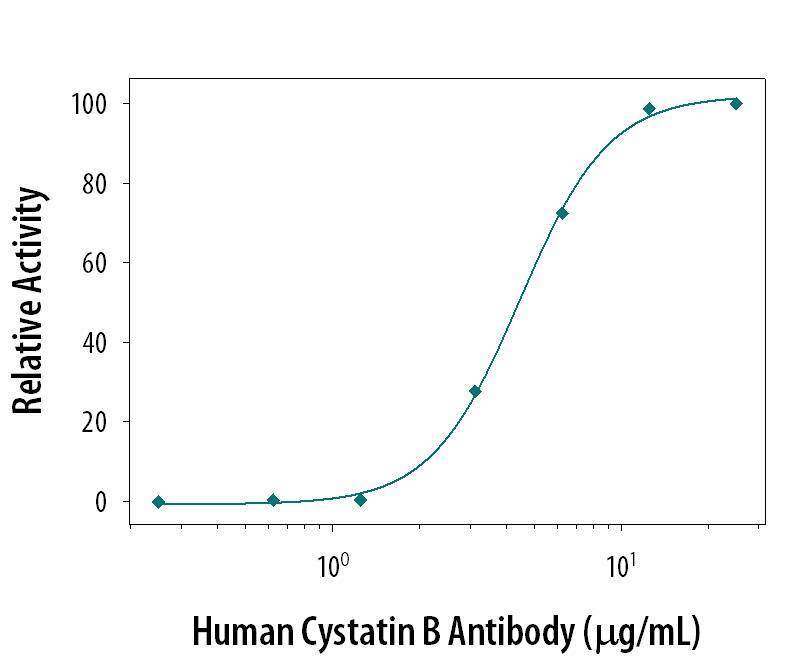
Papain (0.1 µg/mL) activity is measured in the presence of Recombinant Human Cystatin B (0.53 µg/mL, Catalog # 1408-PI) that has been preincubated with increasing concentrations of Human Cystatin B Monoclonal Antibody (Catalog # MAB1408). The ND50 is typically 5.4 µg/mL.Western Blot Shows Human Cystatin B Specificity by Using Knockout Cell Line.
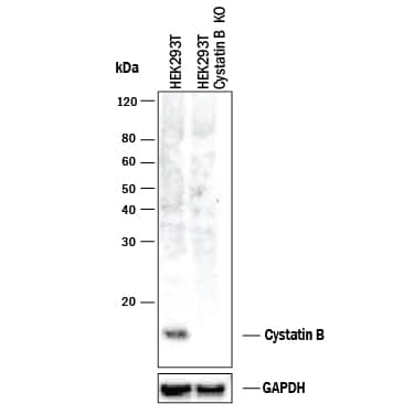
Western blot shows lysates of HEK293T human embryonic kidney parental cell line and Cystatin B knockout HEK293T cell line (KO). PVDF membrane was probed with 2 µg/mL of Mouse Anti-Human Cystatin B Monoclonal Antibody (Catalog # MAB1408) followed by HRP-conjugated Anti-Mouse IgG Secondary Antibody (Catalog # HAF018). A specific band was detected for Cystatin B at approximately 12 kDa (as indicated) in the parental HEK293T cell line, but is not detectable in knockout HEK293T cell line. GAPDH (Catalog # MAB5718) is shown as a loading control. This experiment was conducted under reducing conditions and using Immunoblot Buffer Group 1.Applications for Human Cystatin B Antibody
Immunoprecipitation
Sample: Conditioned cell culture medium spiked with Recombinant Human Cystatin B (Catalog # 1408-PI), see our available Western blot detection antibodies
Knockout Validated
Western Blot
Sample: Human colon tissue, SK‑BR‑3 human breast cancer cell line, and A549 human lung carcinoma cell line
Formulation, Preparation, and Storage
Purification
Reconstitution
Formulation
Shipping
Stability & Storage
- 12 months from date of receipt, -20 to -70 °C as supplied.
- 1 month, 2 to 8 °C under sterile conditions after reconstitution.
- 6 months, -20 to -70 °C under sterile conditions after reconstitution.
Background: Cystatin B
Cystatin B, also called stefin B or liver thiol proteinase inhibitor, is a member of family 1 of the cystatin superfamily (1). Like Cystatin A, it is an intracellular inhibitor regulating the activities of cysteine proteases of the papain family such as cathepsins B, H and L (2). Mutations in the Cystatin B gene is the cause of progressive myoclonus epilepsy (EPM1) (3). Because of its expression patterns, Cystatin B can be used as a marker for certain cancers, such as glioblastoma tumors (4). It readily forms amyloid fibrils in vitro (5). The human Cystatin B consists of 98 amino acid residues (3).
References
- Abrahamson, M. (1994) Methods Enzymol. 244:685.
- Pol, E. and I. Bjork (1999) Biochemistry 38:10519.
- Pennacchio, L.A. et al. (1996) Science 271:1731.
- Zhang, R. et al. (2003) Glia 42:194.
- Zerovnik, E. et al. (2002) Biochim. Biophys. Acta 1594:1.
Alternate Names
Gene Symbol
UniProt
Additional Cystatin B Products
Product Documents for Human Cystatin B Antibody
Product Specific Notices for Human Cystatin B Antibody
For research use only


