Human Desmoglein-2 Antibody
R&D Systems, part of Bio-Techne | Catalog # AF947


Key Product Details
Species Reactivity
Validated:
Cited:
Applications
Validated:
Cited:
Label
Antibody Source
Product Specifications
Immunogen
Met1-Gly608
Accession # CAA81226
Specificity
Clonality
Host
Isotype
Scientific Data Images for Human Desmoglein-2 Antibody
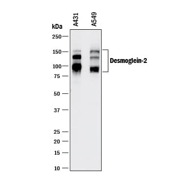
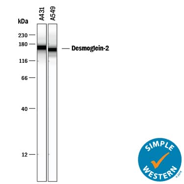
Detection of Human Desmoglein‑2 by Western Blot.
Western blot shows lysates of A431 human epithelial carcinoma cell line and A549 human lung carcinoma cell line. PVDF membrane was probed with 0.25 µg/mL of Goat Anti-Human Desmoglein-2 Antigen Affinity-purified Polyclonal Antibody (Catalog # AF947) followed by HRP-conjugated Anti-Goat IgG Secondary Antibody (Catalog # HAF017). Specific bands were detected for Desmoglein-2 at approximately 90-160 kDa (as indicated). This experiment was conducted under reducing conditions and using Immunoblot Buffer Group 1.Detection of Human Desmoglein‑2 by Simple WesternTM.
Simple Western lane view shows lysates of A431 human epithelial carcinoma cell line and A549 human lung carcinoma cell line, loaded at 0.2 mg/mL. A specific band was detected for Desmoglein-2 at approximately 159-164 kDa (as indicated) using 5 µg/mL of Goat Anti-Human Desmoglein-2 Antigen Affinity-purified Polyclonal Antibody (Catalog # AF947) followed by 1:50 dilution of HRP-conjugated Anti-Goat IgG Secondary Antibody (Catalog # HAF109). This experiment was conducted under reducing conditions and using the 12-230 kDa separation system.Applications for Human Desmoglein-2 Antibody
Simple Western
Sample: A431 human epithelial carcinoma cell line and A549 human lung carcinoma cell line
Western Blot
Sample: A431 human epithelial carcinoma cell line and A549 human lung carcinoma cell line
Formulation, Preparation, and Storage
Purification
Reconstitution
Formulation
Shipping
Stability & Storage
- 12 months from date of receipt, -20 to -70 °C as supplied.
- 1 month, 2 to 8 °C under sterile conditions after reconstitution.
- 6 months, -20 to -70 °C under sterile conditions after reconstitution.
Background: Desmoglein-2
Desmoglein-2 is one of three members of the desmoglein subfamily of calcium-dependent cadherin cell adhesion molecules. Together with desmocollins, another subfamily within the cadherin superfamily, the desmoglein isoforms form the adhesive components of desmosomes, the cell-cell adhesive structures that are found in epithelial cells. Human Desmoglein-2 is a type I transmembrane glycoprotein of 1117 amino acid (aa) residues with a 23 aa signal peptide and a 25 aa propeptide. It differs from other classic cadherins by having four instead of five cadherin repeat domains in its extracellular region, and a much larger cytoplasmic region containing five desmoglein repeat domains which share homology with the cadherin repeats. Instead of having the HAV adhesion motif found in type I cadherins, desmogleins have R/YAL as the adhesion motif on its amino-terminal cadherin repeat. The cytoplasmic tails of desmogleins interact with desmoplakins, plakoglobin and plakophilins. In turn, these proteins link the desmogleins with the intermediate filaments. Desmoglein-2 has been shown to be important in establishing cell-cell adhesion and function in epithelial cells. Desmoglein-2 was originally identified in colon carcinoma and colon, and was named HDGC (human desmoglein colon).
References
- Nollet, R. et al. (2000) J. Mol. Biol. 299:551.
- Elias, P. et al. (2001) J. Cell Biol. 153:243.
- Arnemann, J. et al. (1992) Genomics 13:484.
Alternate Names
Gene Symbol
UniProt
Additional Desmoglein-2 Products
Product Documents for Human Desmoglein-2 Antibody
Product Specific Notices for Human Desmoglein-2 Antibody
For research use only
