Human DISC1 Antibody
R&D Systems, part of Bio-Techne | Catalog # AF6699

Key Product Details
Species Reactivity
Validated:
Cited:
Applications
Validated:
Cited:
Label
Antibody Source
Product Specifications
Immunogen
Lys101-Arg260
Accession # Q9NRI5
Specificity
Clonality
Host
Isotype
Scientific Data Images for Human DISC1 Antibody
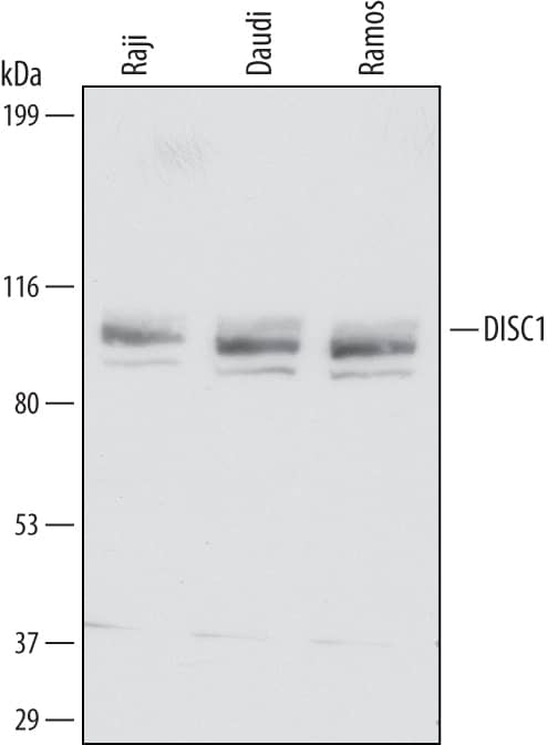
Detection of Human DISC1 by Western Blot.
Western blot shows lysates of Raji human Burkitt's lymphoma cell line, Daudi human Burkitt's lymphoma cell line, and Ramos human Burkitt's lymphoma cell line. PVDF Membrane was probed with 1 µg/mL of Sheep Anti-Human DISC1 Antigen Affinity-purified Polyclonal Antibody (Catalog # AF6699) followed by HRP-conjugated Anti-Sheep IgG Secondary Antibody (Catalog # HAF016). A specific band was detected for DISC1 at approximately 100-105 kDa (as indicated). This experiment was conducted under reducing conditions and using Immunoblot Buffer Group 1.DISC1 in Human Brain.
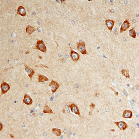
DISC1 was detected in immersion fixed paraffin-embedded sections of human brain (hippocampus) using Sheep Anti-Human DISC1 Antigen Affinity-purified Polyclonal Antibody (Catalog # AF6699) at 10 µg/mL overnight at 4 °C. Before incubation with the primary antibody, tissue was subjected to heat-induced epitope retrieval using Antigen Retrieval Reagent-Basic (Catalog # CTS013). Tissue was stained using the Anti-Sheep HRP-DAB Cell & Tissue Staining Kit (brown; Catalog # CTS019) and counterstained with hematoxylin (blue). Specific staining was localized to neuronal cytoplasm. View our protocol for Chromogenic IHC Staining of Paraffin-embedded Tissue Sections.Applications for Human DISC1 Antibody
Immunohistochemistry
Sample: Immersion fixed paraffin-embedded sections of human brain (hippocampus)
Western Blot
Sample: Raji human Burkitt's lymphoma cell line, Daudi human Burkitt's lymphoma cell line, and Ramos human Burkitt's lymphoma cell line
Formulation, Preparation, and Storage
Purification
Reconstitution
Formulation
Shipping
Stability & Storage
- 12 months from date of receipt, -20 to -70 °C as supplied.
- 1 month, 2 to 8 °C under sterile conditions after reconstitution.
- 6 months, -20 to -70 °C under sterile conditions after reconstitution.
Background: DISC1
DISC1 (Disrupted in Schizophrenia 1) is a 100-105 kDa cytoplasmic and mitochondrial protein that belongs to no known molecular family. It is widely expressed, and appears to have multiple interaction partners, among which are NDEL1, alpha-tubulin, TRAF3IP1 and GSK-3 beta. DISC1 is of particular interest in the brain where it appears to play a role in both neuronal proliferation and migration. Regarding proliferation, DISC1 inhibits GSK-3 beta activity, resulting in neural progenitor cell proliferation without differentiation. With respect to migration, DISC1 promotes embryonic subventricular neuron migration while inhibiting widespread adult neuronal migration from the hippocampal subgranular layer. Human DISC1 is 854 amino acids (aa) in length. It contains an N-terminal globular domain (aa 1-346) plus four coiled-coil regions (aa 366‑830). DISC1 is phosphorylated and forms homodimers. There are multiple isoforms. Among them is a 65-70 kDa form that shows an 18 aa substitution for aa 661-854, a 48 kDa form that contains a 20 aa substitution for aa 350-854, a 61-64 kDa form that possesses a seven aa substitution for aa 545-854, and a 90 kDa form that contains a deletion of aa 748-769. Over aa 101-260, human DISC1 shares 44% aa identity with mouse DISC1.
Long Name
Alternate Names
Gene Symbol
UniProt
Additional DISC1 Products
Product Documents for Human DISC1 Antibody
Product Specific Notices for Human DISC1 Antibody
For research use only

