Human Dkk-3 Antibody
R&D Systems, part of Bio-Techne | Catalog # AF1118

Key Product Details
Species Reactivity
Validated:
Cited:
Applications
Validated:
Cited:
Label
Antibody Source
Product Specifications
Immunogen
Ala22-Ile350
Accession # Q9UBP4
Specificity
Clonality
Host
Isotype
Scientific Data Images for Human Dkk-3 Antibody
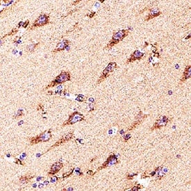
Detection of Dkk-3 in Human Brain Hippocampus.
Dkk-3 was detected in immersion fixed paraffin-embedded sections of Human Brain Hippocampus using Goat Anti-Human Dkk-3 Antigen Affinity-purified Polyclonal Antibody (Catalog # AF1118) at 3 µg/mL for 1 hour at room temperature followed by incubation with the Anti-Goat IgG VisUCyte™ HRP Polymer Antibody (Catalog # VC004). Before incubation with the primary antibody, tissue was subjected to heat-induced epitope retrieval using VisUCyte Antigen Retrieval Reagent-Basic (Catalog # VCTS021). Tissue was stained using DAB (brown) and counterstained with hematoxylin (blue). Specific staining was localized to cytoplasm in neurons. View our protocol for IHC Staining with VisUCyte HRP Polymer Detection Reagents.Detection of Human Dkk-3 by Immunohistochemistry
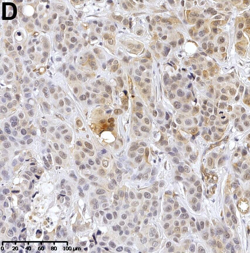
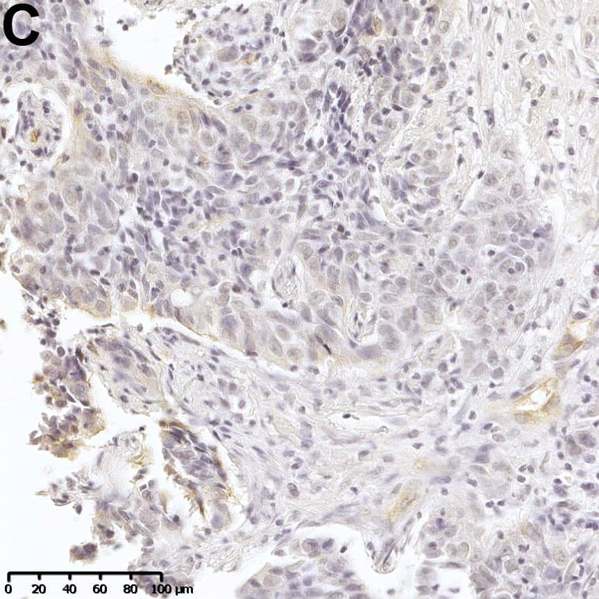
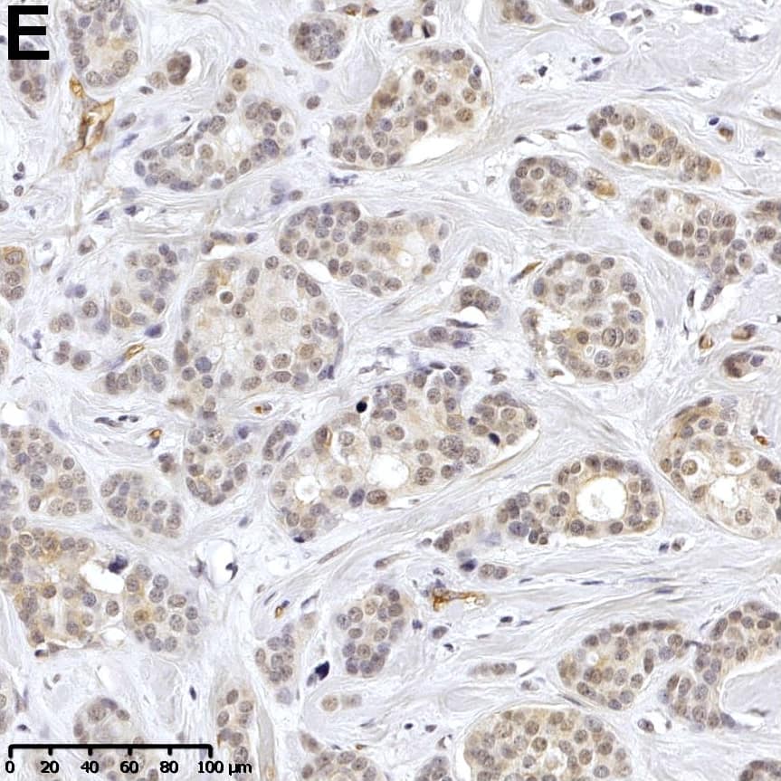
Loss of DKK3 protein expression in human breast cancer.(A) Normal mammary epithelial cells showing moderate, predominantly cytoplasmic, DKK3 immunoreactivity whereas (B) primary antibody negative control is free of signal. (C-E) Weak DKK3 protein expression is observed in breast tumor samples, with lowest intensity in IHC-defined (C) TNBC cases compared to (D) HER2-positive and (E) luminal carcinomas (representative images). (F) Box plot analysis demonstrating a significant down-regulation of DKK3 in tumors of the TNBC (n = 54), the HER2-positive (n = 47) and luminal subtype (n = 362) compared to normal breast tissues (n = 11). Horizontal lines: grouped medians. Boxes: 25-75% quartiles. Vertical lines: range, minimum and maximum. * P < 0.05, *** P < 0.001, IRS: immunoreactive score. Image collected and cropped by CiteAb from the following open publication (https://pubmed.ncbi.nlm.nih.gov/27467270), licensed under a CC-BY license. Not internally tested by R&D Systems.Detection of Human Dkk-3 by Western Blot
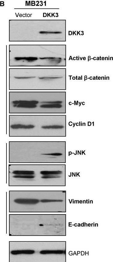
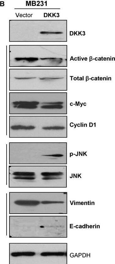
Ectopic expression of DKK3 in MB231 cells disrupted Wnt signalling. (A) Subcellular localization of beta-catenin and active-beta -catenin by immunofluorescence staining. (B) Western blot analysis of beta-catenin, its downstream targets, JNK and EMT markers. (C) RT-PCR analysis of representative stem cell markers in DKK3-transfected MB231 cells. *Indicates significantly down-regulated bands. Image collected and cropped by CiteAb from the following open publication (https://pubmed.ncbi.nlm.nih.gov/23890219), licensed under a CC-BY license. Not internally tested by R&D Systems.Applications for Human Dkk-3 Antibody
ELISA
This antibody functions as an ELISA detection antibody when paired with Mouse Anti-Human Dkk-3 Monoclonal Antibody (Catalog # MAB11181).
This product is intended for assay development on various assay platforms requiring antibody pairs.
Immunohistochemistry
Sample: immersion fixed paraffin-embedded sections of Human Brain Hippocampus
Formulation, Preparation, and Storage
Purification
Reconstitution
Formulation
Shipping
Stability & Storage
- 12 months from date of receipt, -20 to -70 °C as supplied.
- 1 month, 2 to 8 °C under sterile conditions after reconstitution.
- 6 months, -20 to -70 °C under sterile conditions after reconstitution.
Background: Dkk-3
Dkk-3, also known as REIC (Reduced Expansion in Immortalized Cells), is one of four numbered members of the Dickkopf family of Wnt antagonists (1). Dkk-3 is a secreted monomer expressed in many normal human tissues, most strongly in heart, brain and spinal cord (1, 2), and during early embryonic development in the mouse (3). N-glycosylation at up to four sites preceding or between two conserved cysteine-rich motifs results in expression of a 38 kDa glycoprotein (1, 4). The cysteine-rich motifs contain 10 cysteines each, with prokineticin and colipase families containing sequences similar to those of the second motif (1, 5). Human Dkk-3 shows 82%, 88%, 85%, and 53% amino acid (aa) identity with mouse, bovine, canine, and chick Dkk-3, respectively, and 37-45% aa identity with other human Dkk family members. Several lines of evidence implicate Dkk-3 as a negative growth regulator. Dkk-3 is downregulated in many tumors as compared to normal cells, sometimes by loss of heterozygosity (4, 6). Downregulation by CpG hypermethylation in acute lymphoblastic leukemia is correlated with faster progression and shorter survival (7). Release of cultured cells from serum starvation results in downregulation of Dkk-3 in late G1 phase of the cell cycle (6). Over-expression of
Dkk‑3 results in tumor cell-line-specific growth inhibition, induction of apoptosis, and decreased tumorigenicity in nude mice (2, 4, 6). The prototype Dickkopf member, Dkk-1, antagonizes Wnt family signaling by binding to Wnt receptors LRP5 and LRP6 (low-density lipoprotein receptor-related proteins) and promoting their internalization (1, 9, 10). Results are less straightforward for Dkk-3, where some studies show binding to LRP5/6 while others do not. These effects appear to be dependent on the cells and conditions used (1, 6-10).
References
- Krupnik, V.E. et al. (1999) Gene 238:301.
- Tsuji, T. et al. (2000) Biochem. Biophys. Res. Comm. 268:20.
- Kemp, C. et al. (2005) Dev. Dyn. 233:1064.
- Hsieh, S.-Y. et al. (2004) Oncogene 23:9183.
- Bullock, C.M. et al. (2004) Mol. Pharmacol. 65:582.
- Tsuji, T. et al. (2001) Biochem. Biophys. Res. Comm. 289:257.
- Roman-Gomez, J. et al. (2004) Br. J. Cancer 91:707.
- Hoang, B.H. et al. (2004) Cancer Res. 64:2734.
- Caricasole, A. et al. (2003) J. Biol. Chem. 278:37024.
- Mao, B. et al. (2001) Nature 411:321.
Long Name
Alternate Names
Gene Symbol
UniProt
Additional Dkk-3 Products
Product Documents for Human Dkk-3 Antibody
Product Specific Notices for Human Dkk-3 Antibody
For research use only
