Human Dopamine D2 R/DRD2 Antibody
R&D Systems, part of Bio-Techne | Catalog # MAB9266

Key Product Details
Species Reactivity
Validated:
Cited:
Applications
Validated:
Cited:
Label
Antibody Source
Product Specifications
Immunogen
Accession # P14416
Specificity
Clonality
Host
Isotype
Scientific Data Images for Human Dopamine D2 R/DRD2 Antibody
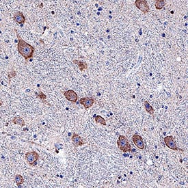
Dopamine D2 R/DRD2 in Human Brain.
Dopamine D2 R/DRD2 was detected in immersion fixed paraffin-embedded sections of human brain (striatum) using Mouse Anti-Human Dopamine D2 R/DRD2 Monoclonal Antibody (Catalog # MAB9266) at 5 µg/mL for 1 hour at room temperature followed by incubation with the Anti-Mouse IgG VisUCyte™ HRP Polymer Antibody (Catalog # VC001). Tissue was stained using DAB (brown) and counterstained with hematoxylin (blue). Specific staining was localized to neuronal cell membranes and cytoplasm. View our protocol for Chromogenic IHC Staining of Paraffin-embedded Tissue Sections.Detection of Human Dopamine D2R/DRD2 by Immunocytochemistry/ Immunofluorescence
Expression of MCT8 and OATP1C1 in D2-MSN (indirect pathway medium-sized spiny neurons) in human and macaque striatum. Representative confocal microscope compositions from double-stained sections for MCT8 (green) or OATP1C1 (green) and for the D2-MSN marker DRD2 (red) in the putamen. Merged images (right side) show the colocalization of the two signals. Coexpression of MCT8 and DRD2 is observed in human (A–C) and macaque (D–F) striatum. Coexpression of OATP1C1 and DRD2 is observed in human (G–I) and macaque (J–L) striatum. Counterstaining with DAPI (blue) shows nuclei of all cells. Note that in humans, the MCT8 signals are located mainly at the cell membrane, while in macaques, they are also located in the cytoplasm. D2-MSN: D2 receptor-expressing medium-sized spiny neurons; DRD2: Dopamine receptor type 2; Put: putamen. Scale bar = 50 μm. Image collected and cropped by CiteAb from the following open publication (https://pubmed.ncbi.nlm.nih.gov/37298594), licensed under a CC-BY license. Not internally tested by R&D Systems.Applications for Human Dopamine D2 R/DRD2 Antibody
Immunohistochemistry
Sample: Immersion fixed paraffin-embedded sections of human brain (striatum)
Formulation, Preparation, and Storage
Purification
Reconstitution
Formulation
Shipping
Stability & Storage
- 12 months from date of receipt, -20 to -70 °C as supplied.
- 1 month, 2 to 8 °C under sterile conditions after reconstitution.
- 6 months, -20 to -70 °C under sterile conditions after reconstitution.
Background: Dopamine D2 R/DRD2
Dopamine receptor D2 (DRD2) is localized to human striatum, motor cortex and neocortex. It is a highly conserved seven transmembrane receptor member of the G-protein coupled receptor 1 family, with three known isoforms. In the striatum, DRD2 suppresses voluntary activity in the striatopallidal pathway, and polymorphisms in this gene are associated with alcohol addiction, smoking behavior, schizophrenia, food addiction and post-traumatic stress disorder as well as myoclonus dystonia. DRD2 has been shown to heterodimerize with DDR4 and 5-HT2A receptors, and traffics between the plasma membrane and intracellular pools and this localization can be modulated by several drugs. DRD2 is localized to human striatum, motor cortex and neocortex.
Long Name
Alternate Names
Gene Symbol
UniProt
Additional Dopamine D2 R/DRD2 Products
Product Documents for Human Dopamine D2 R/DRD2 Antibody
Product Specific Notices for Human Dopamine D2 R/DRD2 Antibody
For research use only

