Human EMMPRIN/CD147 Antibody
R&D Systems, part of Bio-Techne | Catalog # MAB9721

Key Product Details
Validated by
Knockout/Knockdown
Species Reactivity
Human
Applications
Immunocytochemistry, Immunohistochemistry, Knockout Validated, Western Blot
Label
Unconjugated
Antibody Source
Monoclonal Mouse IgG2B Clone # 1036020
Product Specifications
Immunogen
Human embryonic kidney cell HEK293-derived human EMMPRIN/CD147 protein
Glu138-Ala323
Accession # P35613.2
Glu138-Ala323
Accession # P35613.2
Specificity
Detects human EMMPRIN/CD147 in direct ELISAs and Western blots.
Clonality
Monoclonal
Host
Mouse
Isotype
IgG2B
Scientific Data Images for Human EMMPRIN/CD147 Antibody
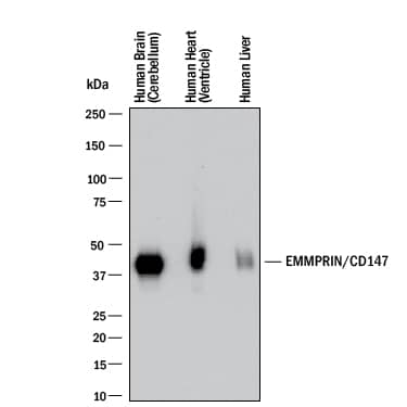
Detection of Human EMMPRIN/CD147 by Western Blot.
Western blot shows lysates of human brain (cerebellum), human heart (ventricle), and human liver. PVDF membrane was probed with 2 µg/mL of Mouse Anti-Human EMMPRIN/CD147 Monoclonal Antibody (Catalog # MAB9721) followed by HRP-conjugated Anti-Mouse IgG Secondary Antibody (HAF018). A specific band was detected for EMMPRIN/CD147 at approximately 45-60 kDa (as indicated). This experiment was conducted under reducing conditions and using Western Blot Buffer Group 1.EMMPRIN/CD147 in 789-O Human Cell Line.
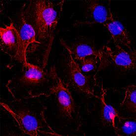
EMMPRIN/CD147 was detected in immersion fixed 789-O Human renal cell carcinoma cell line using Mouse Anti-Human EMMPRIN/CD147 Monoclonal Antibody (Catalog # MAB9721) at 8 µg/mL for 3 hours at room temperature. Cells were stained using the NorthernLights™ 557-conjugated Anti-Mouse IgG Secondary Antibody (red; NL007) and counterstained with DAPI (blue). Specific staining was localized to cell membrane. Staining was performed using our protocol for Fluorescent ICC Staining of Non-adherent Cells.EMMPRIN/CD147 in Human Heart.
EMMPRIN/CD147 was detected in immersion fixed paraffin-embedded sections of human heart using Mouse Anti-Human EMMPRIN/CD147 Monoclonal Antibody (Catalog # MAB9721) at 5 µg/mL for 1 hour at room temperature followed by incubation with the Anti-Mouse IgG VisUCyte™ HRP Polymer Antibody (VC001). Before incubation with the primary antibody, tissue was subjected to heat-induced epitope retrieval using Antigen Retrieval Reagent-Basic (CTS013). Tissue was stained using DAB (brown) and counterstained with hematoxylin (blue). Specific staining was localized to cell membrane in capillaries. Staining was performed using our protocol for IHC Staining with VisUCyte HRP Polymer Detection Reagents.Applications for Human EMMPRIN/CD147 Antibody
Application
Recommended Usage
Immunocytochemistry
8-25 µg/mL
Sample: Immersion fixed 789-O Human renal cell carcinoma cell line
Sample: Immersion fixed 789-O Human renal cell carcinoma cell line
Immunohistochemistry
5-25 µg/mL
Sample: Immersion fixed paraffin-embedded sections of human heart
Sample: Immersion fixed paraffin-embedded sections of human heart
Knockout Validated
2 µg/mL
Sample: EMMPRIN/CD147 is specifically detected in HEK293T human embryonic kidney parental cell line but is not detectable in EMMPRIN/CD147 knockout HEK293T human embryonic kidney cell line.
Sample: EMMPRIN/CD147 is specifically detected in HEK293T human embryonic kidney parental cell line but is not detectable in EMMPRIN/CD147 knockout HEK293T human embryonic kidney cell line.
Western Blot
2 µg/mL
Sample: Human brain (cerebellum), human heart (ventricle), and human liver
Sample: Human brain (cerebellum), human heart (ventricle), and human liver
Reviewed Applications
Read 1 review rated 5 using MAB9721 in the following applications:
Formulation, Preparation, and Storage
Purification
Protein A or G purified from hybridoma culture supernatant
Reconstitution
Reconstitute at 0.5 mg/mL in sterile PBS. For liquid material, refer to CoA for concentration.
Formulation
Lyophilized from a 0.2 μm filtered solution in PBS with Trehalose. *Small pack size (SP) is supplied either lyophilized or as a 0.2 µm filtered solution in PBS.
Shipping
Lyophilized product is shipped at ambient temperature. Liquid small pack size (-SP) is shipped with polar packs. Upon receipt, store immediately at the temperature recommended below.
Stability & Storage
Use a manual defrost freezer and avoid repeated freeze-thaw cycles.
- 12 months from date of receipt, -20 to -70 °C as supplied.
- 1 month, 2 to 8 °C under sterile conditions after reconstitution.
- 6 months, -20 to -70 °C under sterile conditions after reconstitution.
Background: EMMPRIN/CD147
References
- Gabison, E. E. et al. (2005) Biochimie 87:361.
- Yurchenko, V. et al. (2006) Immunology 117:301.
- Kasinrerk, W. et al. (1992) J. Immunol. 149:847.
- Iacono, K.T. et al. (2007) Exp. Mol. Pathol. 83:283.
- Hanna, S. M. et al. (2003) BMC Biochem. 4:17.
- Liao, C-G. et al. (2011) Mol. Cell. Biol. 31:2591.
- Riethdorf, S. et al. (2006) Int. J. Cancer 119:1800.
- Braundmeier, A. G. et al. (2006) J. Clin. Endocrinol. Metab. 91:2358.
- Tang, Y. et al. (2006) Mol. Cancer Res. 4:371.
- Quemener, C. et al. (2007) Cancer Res. 67:9.
- Wilson, M. C. et al. (2005) J. Biol. Chem. 280:27213.
- Xu, D. and M. E. Hemler, (2005) Mol. Cell. Proteomics 4:1061.
- Tang, W. et al. (2004) Mol. Biol. Cell 15:4043.
- Zhao, P. et al. (2010) Cancer Sci. 101:387.
- Dai, J. et al. (2009) BMC Cancer 9:337.
- Li, Y. et al. (2012) J. Biol. Chem. 287:4759.
- Hibino, T. et al. (2013) Cancer Res. 73:172.
- Arora, K. et al. (2005) J. Immunol. 175:517.
- Pushkarsky, T. et al. (2005) J. Biol. Chem. 280:27866.
- Egawa, N. et al. (2006) J. Biol. Chem. 281:37576.
- Sidhu, S. S. et al. (2004) Oncogene 23:956.
- Ulrich, H. et al. (2020) Stem Cell Rev. and Rep., https://doi.org/10.1007/s12015-020-09976-7.
Long Name
Extracellular Matrix Metalloproteinase Inducer
Alternate Names
Basigin, BSG, CD147
Gene Symbol
BSG
UniProt
Additional EMMPRIN/CD147 Products
Product Documents for Human EMMPRIN/CD147 Antibody
Product Specific Notices for Human EMMPRIN/CD147 Antibody
For research use only
Loading...
Loading...
Loading...
Loading...


