Human Fc gamma RIII (CD16) Antibody
R&D Systems, part of Bio-Techne | Catalog # MAB4325


Key Product Details
Species Reactivity
Validated:
Cited:
Applications
Validated:
Cited:
Label
Antibody Source
Product Specifications
Immunogen
Gly17-Gln208
Accession # P08637
Specificity
Clonality
Host
Isotype
Scientific Data Images for Human Fc gamma RIII (CD16) Antibody
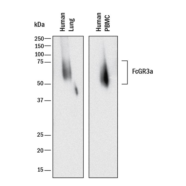
Detection of Human Fc gamma RIII (CD16) by Western Blot.
Western blot shows lysates of human lung tissue and human peripheral blood mononuclear cells (PBMCs). PVDF membrane was probed with 2 µg/mL of Mouse Anti-Human Fc gamma RIII (CD16) Monoclonal Antibody (Catalog # MAB4325) followed by HRP-conjugated Anti-Mouse IgG Secondary Antibody (Catalog # HAF018). A specific band was detected for Fc gamma RIII (CD16) at approximately 50 kDa (as indicated). This experiment was conducted under reducing conditions and using Immunoblot Buffer Group 1.Detection of Fc gamma RIII (CD16) in Human PBMCs by Flow Cytometry.
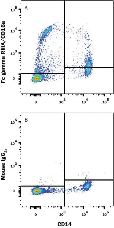
Human peripheral blood mononuclear cells (PBMCs) were stained with (A) Mouse Anti-Human Fc gamma RIII (CD16) Monoclonal Antibody (Catalog # MAB4325) or (B) Mouse IgG2A isotype control antibody (Catalog # MAB003) followed by Goat anti-Mouse IgG APC-conjugated Secondary Antibody (Catalog # F0101B) and Mouse anti-Human CD14 PE-conjugated Monoclonal Antibody (Catalog # FAB3832P). View our protocol for Staining Membrane-associated Proteins.Fc gamma RIII (CD16) in Human PBMCs.
Fc gamma RIII (CD16) was detected in immersion fixed human peripheral blood mononuclear cells (PBMCs) using Mouse Anti-Human Fc gamma RIII (CD16) Monoclonal Antibody (Catalog # MAB4325) at 8 µg/mL for 3 hours at room temperature. Cells were stained using the NorthernLights™ 557-conjugated Anti-Mouse IgG Secondary Antibody (red; Catalog # NL007) and counterstained with DAPI (blue). Specific staining was localized to cell surfaces. View our protocol for Fluorescent ICC Staining of Non-adherent Cells.Applications for Human Fc gamma RIII (CD16) Antibody
CyTOF-ready
Flow Cytometry
Sample: Human PBMC
Immunocytochemistry
Sample: Immersion fixed human peripheral blood mononuclear cells (PBMCs)
Immunohistochemistry
Sample: Immersion fixed paraffin-embedded sections of human spleen
Western Blot
Sample: Human lung tissue and Human peripheral blood mononuclear cells (PBMCs)
Formulation, Preparation, and Storage
Purification
Reconstitution
Formulation
Shipping
Stability & Storage
- 12 months from date of receipt, -20 to -70 °C as supplied.
- 1 month, 2 to 8 °C under sterile conditions after reconstitution.
- 6 months, -20 to -70 °C under sterile conditions after reconstitution.
Background: Fc gamma RIII (CD16)
Fc gamma RIIIa is a low/intermediate affinity receptor for polyvalent immune-complexed IgG. It is involved in phagocytosis, secretion of enzymes and inflammatory mediators, antibody-dependent cytotoxicity and clearance of immune complexes (1, 2). In humans, it is a 50-70 kDa type I transmembrane activating receptor expressed by NK cells, T cells, monocytes, and macrophages (1). Fc gamma RIIIb is highly related, sharing 97% amino acid (aa) identity within the extracellular domain (ECD), but is a GPI-linked receptor expressed on human neutrophils and eosinophils (1, 2). The ECD of Fc gamma RIIIa shares 63%, 61%, 65%, 59% and 58% aa identity with mouse Fc gamma RIV, rat Fc gamma RIIIa, feline CD16, bovine CD16 and porcine Fc gamma RIIIb paralogs, respectively. The Fc gamma RIIIa cDNA encodes 254 aa including a 16 aa signal sequence, 191 aa ECD with two C2-type Ig-like domains and five potential N-glycosylation sites, a 22 aa transmembrane (TM) sequence and a 25 aa cytoplasmic domain. In humans, a single nucleotide polymorphism creates high binding (176V) and low binding (176F) forms that, when homozygous, may influence susceptibility to autoimmune diseases or response to therapeutic IgG antibodies (3, 4). Catalog # 4325-FC is expressed as the 176V isoform of Fc gamma RIIIa. Fc gamma RIIIa surface expression requires interaction of an accessory chain, either the common gamma-chain or CD3 zeta (5, 6). Glycosylation patterns, electrophoretic mobility and binding affinity appear to differ between NK cell and monocyte Fc gamma RIIIa (7). The ECD of both Fc gamma RIIIa and b can be proteolytically cleaved and retain binding activity in soluble form (8-11). In monocytes and macrophages, activation and phagocytosis can trigger Fc gamma RIIIa release (11). Soluble Fc gamma RIII can be detected in normal plasma and is increased in rheumatoid arthritis and in coronary artery diseases (9, 10).
References
- Nimmerjahn, F. and J.V. Ravetch (2006) Immunity 24:19.
- Ravetch, J.V. and B. Perussia (1989) J. Exp. Med. 170:481.
- Wu, J. et al. (1997) J. Clin. Invest. 100:1059.
- Dall’Ozzo, S. et al. (2004) Cancer Res. 64:4664.
- Kim, M.-K. et al. (2003) Blood 101:4479.
- Lanier, L.L. et al. (1989) Nature 342:803.
- Edberg, J.C. and R.P. Kimberley (1997) J. Immunol. 159:3849.
- Li, P. et al. (2007) J. Biol. Chem. 282:6210.
- Masuda, M. et al. (2003) J. Rheumatol. 30:1911.
- Masuda, M. et al. (2006) Atherosclerosis 188:377.
- Webster, N.L. et al. (2006) J. Leukoc. Biol. 79:294.
Long Name
Alternate Names
Gene Symbol
UniProt
Additional Fc gamma RIII (CD16) Products
Product Documents for Human Fc gamma RIII (CD16) Antibody
Product Specific Notices for Human Fc gamma RIII (CD16) Antibody
For research use only


