Human FGFR2 alpha Antibody
R&D Systems, part of Bio-Techne | Catalog # MAB11119

Key Product Details
Species Reactivity
Human
Applications
Flow Cytometry, Immunocytochemistry
Label
Unconjugated
Antibody Source
Monoclonal Mouse IgG2B Clone # 1057909
Product Specifications
Immunogen
Chinese Hamster Ovary cell line, CHO-derived human FGFR2 alpha protein
Arg22-Glu377
Accession # P21802.1
Arg22-Glu377
Accession # P21802.1
Specificity
Detects human FGFR2 alpha in direct ELISA.
Clonality
Monoclonal
Host
Mouse
Isotype
IgG2B
Scientific Data Images for Human FGFR2 alpha Antibody
Detection of FGFR2 alpha in HT-29 (Positive) and HEL 92.1.7 (Negative).
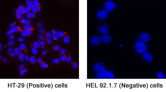
FGFR2 alpha was detected in immersion fixed HT-29 Human Colon Adenocarcinoma Cell Line (Positive) and absent in HEL 92.1.7 Human Erythroleukemic Cell Line (Negative) using Mouse Anti-Human FGFR2 alpha Monoclonal Antibody (Catalog # MAB11119) at 3 µg/mL for 3 hours at room temperature. Cells were stained using the NorthernLights™ 557-conjugated Anti-Mouse IgG Secondary Antibody (red; Catalog # NL007) and counterstained with DAPI (blue). Specific staining was localized to cytoplasm. View our protocol for Fluorescent ICC Staining of Cells on Coverslips.Detection of FGFR2 alpha in KatoIII cells by Flow Cytometry.
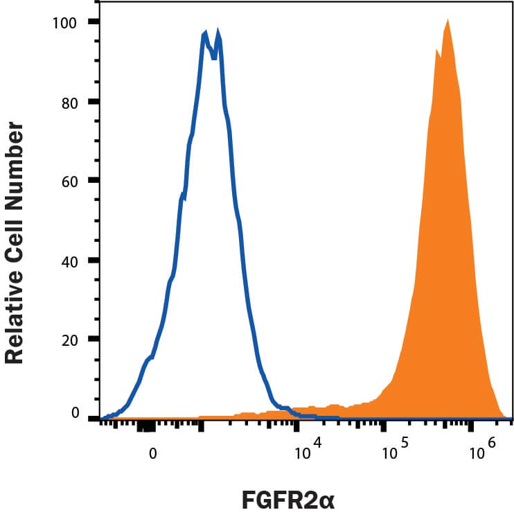
KATO-III human gastric carcinoma cells were stained with Mouse Anti-Human FGFR2 alpha Monoclonal Antibody (Catalog # MAB11119, filled histogram) or isotype control antibody (Catalog # MAB004, open histogram), followed by Allophycocyanin-conjugated Anti-Mouse IgG Secondary Antibody (Catalog # F0101B). View our protocol for Staining Membrane-associated Proteins.Applications for Human FGFR2 alpha Antibody
Application
Recommended Usage
Flow Cytometry
0.25 µg/106 cells
Sample: KATO-III human gastric carcinoma cell line
Sample: KATO-III human gastric carcinoma cell line
Immunocytochemistry
3-25 µg/mL
Sample: Immersion fixed HT-29 Human Colon Adenocarcinoma Cell Line (Positive) and HEL 92.1.7 Human Erythroleukemic Cell Line (Negative)
Sample: Immersion fixed HT-29 Human Colon Adenocarcinoma Cell Line (Positive) and HEL 92.1.7 Human Erythroleukemic Cell Line (Negative)
Formulation, Preparation, and Storage
Reconstitution
Reconstitute at 0.5 mg/mL in sterile PBS. For liquid material, refer to CoA for concentration.
Formulation
Lyophilized from a 0.2 μm filtered solution in PBS with Trehalose. *Small pack size (SP) is supplied either lyophilized or as a 0.2 µm filtered solution in PBS.
Shipping
Lyophilized product is shipped at ambient temperature. Liquid small pack size (-SP) is shipped with polar packs. Upon receipt, store immediately at the temperature recommended below.
Stability & Storage
Use a manual defrost freezer and avoid repeated freeze-thaw cycles.
- 12 months from date of receipt, -20 to -70 °C as supplied.
- 1 month, 2 to 8 °C under sterile conditions after reconstitution.
- 6 months, -20 to -70 °C under sterile conditions after reconstitution.
Background: FGFR2 alpha
References
- Ornitz, D.M. and Itoh, N. (2015) Wiley Interdiscip Rev Dev Biol. 4:215.
- Zhang, X. et al. (2006) J Biol Chem. 281:15694.
- Ferguson, H.R. et al. (2021) Signaling. Cells 10:1201.
- Holzmann, K. et al. (2012) J Nucleic Acids. 2012:950508.
- Wagner, E.J. et al. (2003) RNA 9:1552.
- Xie, Y. et al. (2020) Sig Transduct Target Ther 5:181.
- Mossahebi-Mohammadi, M. et al. (2020) Front Cell Dev Biol. 18:79.
- Teven, C.M. et al. (2014) Genes Dis. 1:199.
- Matsuda, Y. et al. (2012) Mol Cancer Ther. 11:2010.
Long Name
Fibroblast Growth Factor Receptor 2 alpha
Alternate Names
FGF R2a
Gene Symbol
FGFR2
UniProt
Additional FGFR2 alpha Products
Product Documents for Human FGFR2 alpha Antibody
Product Specific Notices for Human FGFR2 alpha Antibody
For research use only
Loading...
Loading...
Loading...
Loading...
Loading...

