Human GluR4 Antibody
R&D Systems, part of Bio-Techne | Catalog # MAB113631


Conjugate
Catalog #
Key Product Details
Species Reactivity
Human
Applications
Flow Cytometry, Immunocytochemistry
Label
Unconjugated
Antibody Source
Monoclonal Mouse IgG2B Clone # 1059705
Product Specifications
Immunogen
Synthetic peptide
Accession # P48058
Accession # P48058
Specificity
Detects human GluR4 in direct ELISA.
Clonality
Monoclonal
Host
Mouse
Isotype
IgG2B
Scientific Data Images for Human GluR4 Antibody
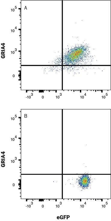
Detection of GluR4 in HEK293 cells transfected with Human GRIA4 and eGFP vs irrelevant cells by Flow Cytometry.
HEK293 cells transfected with Human GRIA4 and eGFP (A) vs irrelevant and eGFP (B) was stained with Mouse Anti-Human GluR4 Monoclonal Antibody (Catalog # MAB113631) followed by Allophycocyanin-conjugated Anti-Mouse IgG Secondary Antibody (Catalog # F0101B). View our protocol for Staining Membrane-associated Proteins.Detection of GluR4 in Transfected HEK293 Cells (Positive) and absent in Wild Type HEK293 Cells (Negative).
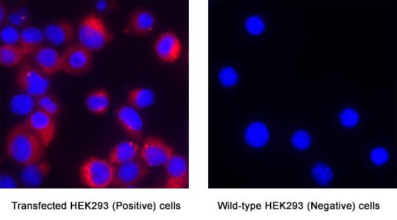
GluR4 was detected in immersion fixed Transfected HEK293 Human Embryonic Kidney Cell Line (Positive) and absent in Wild Type HEK293 Cell Line (Negative) using Mouse Anti-Human GluR4 Monoclonal Antibody (Catalog # MAB113631) at 8 µg/mL for 3 hours at room temperature. Cells were stained using the NorthernLights™ 557-conjugated Anti-Mouse IgG Secondary Antibody (red; Catalog # NL007) and counterstained with DAPI (blue). Specific staining was localized to cytoplasm. View our protocol for Fluorescent ICC Staining of Cells on Coverslips.Applications for Human GluR4 Antibody
Application
Recommended Usage
Flow Cytometry
0.25 µg/106 cells
Sample: GRIA4 HEK293/eGFP and irrelevant HEK293/eGFP transfectants
Sample: GRIA4 HEK293/eGFP and irrelevant HEK293/eGFP transfectants
Immunocytochemistry
5-25 µg/mL
Sample: Immersion fixed Transfected HEK293 Human Embryonic Kidney Cell Line (Positive) and absent in Wild Type HEK293 Human Embryonic Kidney Cell Line (Negative)
Sample: Immersion fixed Transfected HEK293 Human Embryonic Kidney Cell Line (Positive) and absent in Wild Type HEK293 Human Embryonic Kidney Cell Line (Negative)
Formulation, Preparation, and Storage
Purification
Protein A or G purified from cell culture supernatant
Reconstitution
Reconstitute at 0.5 mg/mL in sterile PBS. For liquid material, refer to CoA for concentration.
Formulation
Lyophilized from a 0.2 μm filtered solution in PBS with Trehalose. See Certificate of Analysis for details.
*Small pack size (-SP) is supplied either lyophilized or as a 0.2 µm filtered solution in PBS.
*Small pack size (-SP) is supplied either lyophilized or as a 0.2 µm filtered solution in PBS.
Shipping
Lyophilized product is shipped at ambient temperature. Liquid small pack size (-SP) is shipped with polar packs. Upon receipt, store immediately at the temperature recommended below.
Stability & Storage
Use a manual defrost freezer and avoid repeated freeze-thaw cycles.
- 12 months from date of receipt, -20 to -70 °C as supplied.
- 1 month, 2 to 8 °C under sterile conditions after reconstitution.
- 6 months, -20 to -70 °C under sterile conditions after reconstitution.
Background: GluR4
Long Name
Glutamate Receptor 4
Alternate Names
GluA4, GLURD
Gene Symbol
GRIA4
UniProt
Additional GluR4 Products
Product Documents for Human GluR4 Antibody
Product Specific Notices for Human GluR4 Antibody
For research use only
Loading...
Loading...
Loading...
Loading...
Loading...
