Human HB-EGF Antibody
R&D Systems, part of Bio-Techne | Catalog # AF-259-NA


Key Product Details
Validated by
Species Reactivity
Validated:
Cited:
Applications
Validated:
Cited:
Label
Antibody Source
Product Specifications
Immunogen
Asp63-Leu148
Accession # Q99075
Specificity
Clonality
Host
Isotype
Endotoxin Level
Scientific Data Images for Human HB-EGF Antibody
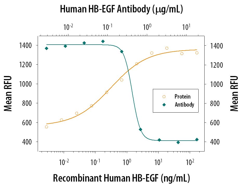
Cell Proliferation Induced by HB‑EGF and Neutralization by Human HB‑EGF Antibody.
Recombinant Human HB-EGF (Catalog # 259-HE) stimulates proliferation in the Balb/3T3 mouse embryonic fibroblast cell line in a dose-dependent manner (orange line). Proliferation elicited by Recombinant Human HB-EGF (20 ng/mL) is neutralized (green line) by increasing concentrations of Human HB-EGF Antigen Affinity-purified Polyclonal Antibody (Catalog # AF-259-NA). The ND50 is typically 0.1-0.6 µg/mL.HB-EGF in Human Placenta.
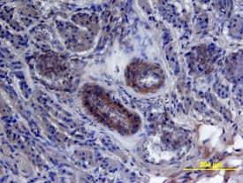
HB-EGF was detected in immersion fixed paraffin-embedded sections of human placenta using 15 µg/mL Human HB-EGF Antigen Affinity-purified Polyclonal Antibody (Catalog # AF-259-NA) overnight at 4 °C. Tissue was stained with the Anti-Goat HRP-DAB Cell & Tissue Staining Kit (brown; Catalog # CTS008) and counterstained with hematoxylin (blue). View our protocol for Chromogenic IHC Staining of Paraffin-embedded Tissue Sections.HB-EGF in Human Placenta.
HB-EGF was detected in immersion fixed paraffin-embedded sections of human placenta using Human HB-EGF Antigen Affinity-purified Polyclonal Antibody (Catalog # AF-259-NA) at 15 µg/mL overnight at 4 °C. Tissue was stained using the Anti-Goat HRP-DAB Cell & Tissue Staining Kit (brown; Catalog # CTS008) and counterstained with hematoxylin (blue). View our protocol for Chromogenic IHC Staining of Paraffin-embedded Tissue Sections.Applications for Human HB-EGF Antibody
Immunohistochemistry
Sample: Immersion fixed paraffin-embedded sections of human placenta
Western Blot
Sample: Recombinant Human HB-EGF (Catalog # 259-HE)
Neutralization
Human HB-EGF Sandwich Immunoassay
Reviewed Applications
Read 1 review rated 4 using AF-259-NA in the following applications:
Formulation, Preparation, and Storage
Purification
Reconstitution
Formulation
Shipping
Stability & Storage
- 12 months from date of receipt, -20 to -70 °C as supplied.
- 1 month, 2 to 8 °C under sterile conditions after reconstitution.
- 6 months, -20 to -70 °C under sterile conditions after reconstitution.
Background: HB-EGF
HB-EGF was originally purified based on its heparin-binding property and mitogenic activity on BALB-3T3 fibroblasts from the conditioned medium of the human U-937 histiocytic lymphoma cell line. The natural protein has an apparent molecular mass of 19-23 kDa and exists in multiple forms as a result of heterogenous O-glycosylation and/or N-terminal truncation. In addition to fibroblasts, HB-EGF is also a potent mitogen for keratinocytes and smooth muscle cells but not for capillary endothelial cells. HB-EGF is produced in monocytes and macrophages. In addition, transcription of HB-EGF can be induced in vascular endothelial cells as well as aortic smooth muscle cells (SMC), suggesting that HB-EGF may have an important role in the pathogenesis of atherosclerosis.
HB-EGF is a member of the EGF family of mitogens which also include transforming growth factor-alpha (TGF-alpha), amphiregulin (AR), rat schwanoma-derived growth factor (SDGF), vaccinia growth factor (VGF), and the various ligands for the Her2/ErbB2/neu receptor. All these cytokines are derived from transmembrane precursors that contain one or several EGF structural units in their extracellular domain. Many of these transmembrane precursors are biologically active and seem to play a role in juxtacrine stimulation of adjacent cells. The cDNA for HB-EGF encodes a 204 amino acid residue transmembrane protein that is proteolytically cleaved to generate the soluble HB-EGF. Like EGF, TGF-alpha, and AR; HB-EGF binds to the EGF receptor and activates the receptor tyrosine kinase. HB-EGF is reported to be a more potent SMC mitogen than EGF. It has been suggested that the differential activities found for HB-EGF compared to EGF may be mediated by the heparin-binding properties of HB-EGF. A diphtheria toxin receptor that mediates the endocytosis of the bound toxin has been cloned and found to be identical to the transmembrane HB-EGF precursor.
Long Name
Alternate Names
Gene Symbol
UniProt
Additional HB-EGF Products
Product Documents for Human HB-EGF Antibody
Product Specific Notices for Human HB-EGF Antibody
For research use only


