Human HIC5/TGFB1I1 Antibody
R&D Systems, part of Bio-Techne | Catalog # AF5626

Key Product Details
Species Reactivity
Applications
Label
Antibody Source
Product Specifications
Immunogen
Val184-Gly288
Accession # O43294
Specificity
Clonality
Host
Isotype
Scientific Data Images for Human HIC5/TGFB1I1 Antibody
Detection of Human HIC5/TGFB1I1 by Western Blot.
Western blot shows lysates of MCF-7 human breast cancer cell line and HepG2 human hepatocellular carcinoma cell line. PVDF membrane was probed with 1 µg/mL of Goat Anti-Human HIC5/TGFB1I1 Antigen Affinity-purified Polyclonal Antibody (Catalog # AF5626) followed by HRP-conjugated Anti-Goat IgG Secondary Antibody (Catalog # HAF109). A specific band was detected for HIC5/TGFB1I1 at approximately 50 kDa (as indicated). This experiment was conducted under reducing conditions and using Immunoblot Buffer Group 1.HIC5/TGFB1I1 in PC‑3 Human Cell Line.
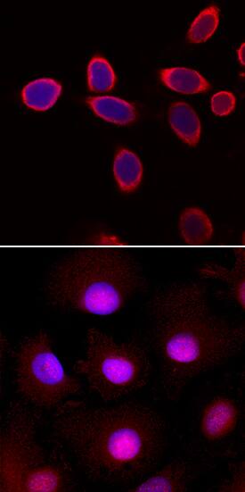
HIC5/TGFB1I1 was detected in immersion fixed PC-3 human prostate cancer cell line, unstimulated (upper panel) or stimulated with 10 ng/mL Recombinant Human BMP-4 (Catalog # 314-BP; lower panel), using Goat Anti-Human HIC5/TGFB1I1 Antigen Affinity-purified Polyclonal Antibody (Catalog # AF5626) at 10 µg/mL for 3 hours at room temperature. Cells were stained using the NorthernLights™ 557-conjugated Anti-Goat IgG Secondary Antibody (red; Catalog # NL001) and counterstained with DAPI (blue). Specific staining was localized to cytoplasm (unstimulated) and nuclei (stimulated). View our protocol for Fluorescent ICC Staining of Cells on Coverslips.Applications for Human HIC5/TGFB1I1 Antibody
Immunocytochemistry
Sample: Immersion fixed PC-3 human prostate cancer cell line stimulated with 10 ng/mL Recombinant Human BMP-4 (Catalog # 314-BP)
Western Blot
Sample: MCF-7 human breast cancer cell line and HepG2 human hepatocellular carcinoma cell line
Formulation, Preparation, and Storage
Purification
Reconstitution
Formulation
Shipping
Stability & Storage
- 12 months from date of receipt, -20 to -70 °C as supplied.
- 1 month, 2 to 8 °C under sterile conditions after reconstitution.
- 6 months, -20 to -70 °C under sterile conditions after reconstitution.
Background: HIC5/TGFB1I1
HIC5 (Hydrogen peroxide inducible clone 5; also ARA55) is a 50-55 kDa group III member of the LIM domain family of proteins. It is expressed primarily in smooth muscle, platelets and myoepithelium. It resides in both cytoplasm and nucleus, and performs multiple functions. It associates with focal adhesions, binds to the nuclear matrix, and serves as a coactivator for the glucocorticoid and androgen receptors. Human HIC5 is 461 amino acids (aa) in length. It contains four Leu:Asp‑rich motifs (aa 1‑215) and four LIM domains (aa 226‑461). LIM domains, either individually, or in combination, perform the majority of functions. LIM4 binds to the nuclear matrix, LIMs 3 and 4 are coactivators, and LIMs 2 and 3 bind to focal adhesions. HIC5 is induced by TGF beta and by hydrogen peroxide.
Long Name
Alternate Names
Gene Symbol
UniProt
Additional HIC5/TGFB1I1 Products
Product Documents for Human HIC5/TGFB1I1 Antibody
Product Specific Notices for Human HIC5/TGFB1I1 Antibody
For research use only

