Human HVEM/TNFRSF14 Antibody
R&D Systems, part of Bio-Techne | Catalog # MAB3563
Recombinant Monoclonal Antibody.


Conjugate
Catalog #
Key Product Details
Species Reactivity
Human
Applications
Flow Cytometry, Neutralization
Label
Unconjugated
Antibody Source
Recombinant Monoclonal Rabbit IgG Clone # 2742B
Product Specifications
Immunogen
Human embryonic kidney cell HEK293-derived human HVEM/TNFRSF14
Pro37-Val202
Accession # Q92956
Pro37-Val202
Accession # Q92956
Specificity
Detects human HVEM/TNFRSF14 in ELISA.
Clonality
Monoclonal
Host
Rabbit
Isotype
IgG
Scientific Data Images for Human HVEM/TNFRSF14 Antibody
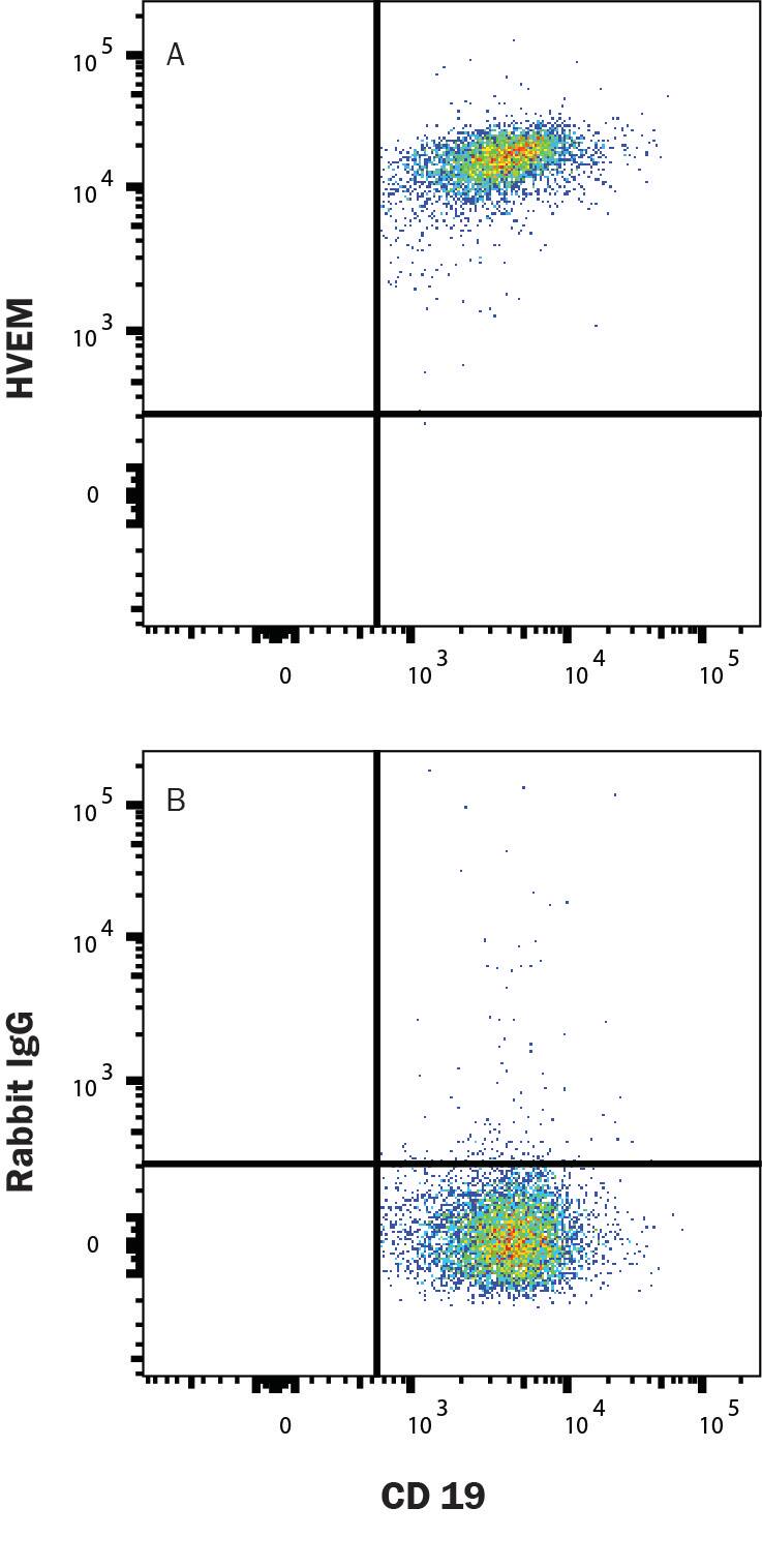
Detection of HVEM/TNFRSF14 in Human PBMC B Cells by Flow Cytometry.
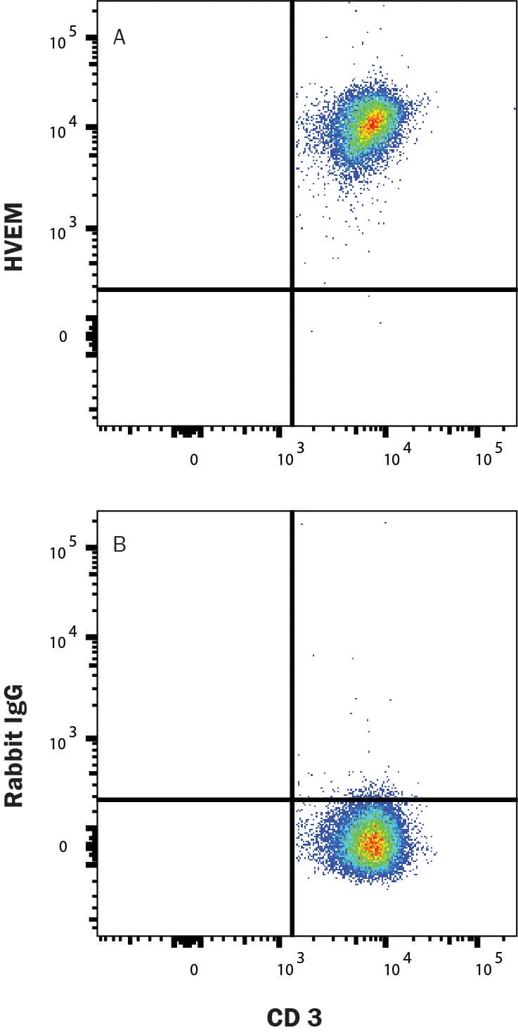
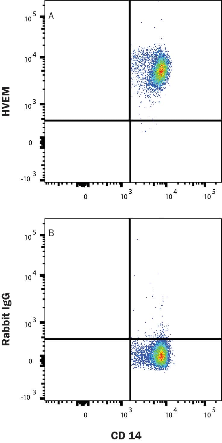
Human PBMC lymphocytes were stained with (A) Rabbit Anti-Human HVEM/TNFRSF14 Monoclonal Antibody (Catalog # MAB3563) or (B) isotype control antibody (MAB1050), followed by Allophycocyanin-conjugated Anti-Rabbit IgG Secondary Antibody (F0111) and Mouse Anti-Human PE-conjugated CD19 (FAB4867P). Cells were gated on Mouse Anti-Human Alexa Fluor® 405-conjugated CD14 (FAB3832V), Mouse Anti-Human Alexa Fluor® 488-conjugated CD3 (FAB100G), and Mouse Anti-Human Alexa Fluor® 700-conjugated CD56 (FAB24086N) negative populations. Staining was performed using our Staining Membrane-associated Proteins protocol.Detection of HVEM/TNFRSF14 in Human PBMC T cells by Flow Cytometry.
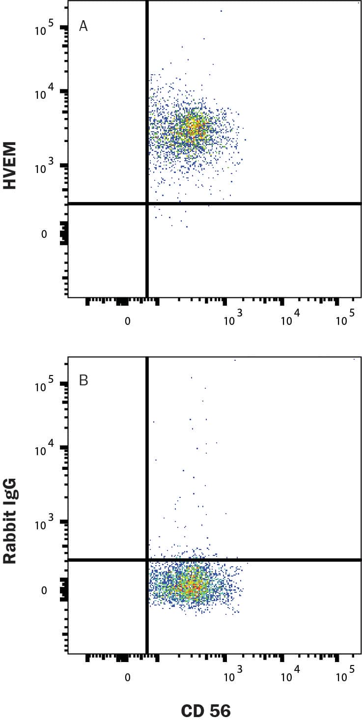
Human PBMC lymphocytes were stained with (A) Rabbit Anti-Human HVEM/TNFRSF14 Monoclonal Antibody (Catalog # MAB3563) or (B) isotype control antibody (MAB1050), followed by Allophycocyanin-conjugated Anti-Rabbit IgG Secondary Antibody (F0111) and Mouse Anti-Human Alexa Fluor® 488-conjugated CD3 (FAB100G). Cells were gated on Mouse Anti-Human Alexa Fluor® 405-conjugated CD14 (FAB3832V), Mouse Anti-Human PE-conjugated CD19 (FAB4867P), and Mouse Anti-Human Alexa Fluor® 700-conjugated CD56 (FAB24086N) negative populations. Staining was performed using our Staining Membrane-associated Proteins protocol.Detection of HVEM/TNFRSF14 in Human PBMC NK cells by Flow Cytometry
Human PBMC lymphocytes were stained with (A) Rabbit Anti-Human HVEM/TNFRSF14 Monoclonal Antibody (Catalog # MAB3563) or (B) isotype control antibody (MAB1050), followed by Allophycocyanin-conjugated Anti-Rabbit IgG Secondary Antibody (F0111) and Mouse Anti-Human Alexa Fluor® 700-conjugated CD56 (FAB24086N). Cells were gated on Mouse Anti-Human Alexa Fluor® 405-conjugated CD14 (FAB3832V), Mouse Anti-Human PE-conjugated CD19 (FAB4867P), and Mouse Anti-Human Alexa Fluor® 488-conjugated CD3 (FAB100G) negative populations. Staining was performed using our Staining Membrane-associated Proteins protocol.Applications for Human HVEM/TNFRSF14 Antibody
Application
Recommended Usage
Flow Cytometry
0.25 µg/106 cells
Sample: Human PBMCs
Sample: Human PBMCs
Neutralization
In a functional ELISA binding assay, 0.075-0.45 ug/mL of this antibody
will block 50% of the binding of 1 ug/mL of
recombinant human BTLA His-tag protein (catalog# 9235-BT) to immobilized
recombinant human HVEM Fc Chimera coat 1 ug/mL (100 µL/well). At 1 ug/mL,
this antibody will block >90% of the binding.
Formulation, Preparation, and Storage
Purification
Protein A or G purified from cell culture supernatant
Reconstitution
Reconstitute at 0.5 mg/mL in sterile PBS. For liquid material, refer to CoA for concentration.
Formulation
Lyophilized from a 0.2 μm filtered solution in PBS with Trehalose. *Small pack size (SP) is supplied either lyophilized or as a 0.2 µm filtered solution in PBS.
Shipping
Lyophilized product is shipped at ambient temperature. Liquid small pack size (-SP) is shipped with polar packs. Upon receipt, store immediately at the temperature recommended below.
Stability & Storage
Use a manual defrost freezer and avoid repeated freeze-thaw cycles.
- 12 months from date of receipt, -20 to -70 °C as supplied.
- 1 month, 2 to 8 °C under sterile conditions after reconstitution.
- 6 months, -20 to -70 °C under sterile conditions after reconstitution.
Background: HVEM/TNFRSF14
References
- del Rio, M.L. et al. (2010) J. Leukoc. Biol. 87:223.
- Montgomery, R.I. et al. (1996) Cell 87:427.
- Hsu, H. et al. (1997) J. Biol. Chem. 272:13471.
- Sedy, J. R. et al. (2005) Nat. Immunol. 6:90.
- Tao, R. et al. (2008) J. Immunol. 180:6649.
- Steinberg, M.W. et al. (2013) PLoS One 8:e77992.
- de Trez, C. et al. (2008) J. Immunol. 180:238.
- Heo, S.K. et al. (2006) J. Leukoc. Biol. 79:330.
- Gonzalez, L.C. et al. (2005) Proc. Natl. Acad Sci. USA 102:1116.
- Cai, G. et al. (2008) Nat. Immunol. 9:176.
- Mauri, D.N. et al. (1998) Immunity 8:21.
- Harrop, J.A. et al. (1998) J. Biol. Chem. 273:27548.
- Wang, J. et al. (2005) J. Immunol. 174:8173.
- Ye, Q. et al. (2002) J. Exp. Med. 195:795.
- Steinberg, M.W. et al. (2008) J. Exp. Med. 205:1463.
- Shui, J.W. et al. (2012) Nature 488:222.
Long Name
Herpesvirus Entry Mediator
Alternate Names
ATAR, CD270, LIGHTR, TNFRSF14
Gene Symbol
TNFRSF14
UniProt
Additional HVEM/TNFRSF14 Products
Product Documents for Human HVEM/TNFRSF14 Antibody
Product Specific Notices for Human HVEM/TNFRSF14 Antibody
For research use only
Loading...
Loading...
Loading...
Loading...
Loading...


