Human ICAM-1/CD54 Fluorescein-conjugated Antibody
R&D Systems, part of Bio-Techne | Catalog # BBA20


Key Product Details
Species Reactivity
Validated:
Human
Cited:
Human
Applications
Validated:
Flow Cytometry
Cited:
Flow Cytometry
Label
Fluorescein (Excitation = 488 nm, Emission = 515-545 nm)
Antibody Source
Monoclonal Mouse IgG1 Clone # BBIG-I1 (11C81)
Product Specifications
Immunogen
Activated HUVEC human umbilical vein endothelial cells
Specificity
Stains human ICAM-1/CD54 on human ICAM-1 transfected COS cells. It does not bind COS cells transfected with E-Selectin, VCAM-1, or PECAM-1.
Clonality
Monoclonal
Host
Mouse
Isotype
IgG1
Scientific Data Images for Human ICAM-1/CD54 Fluorescein-conjugated Antibody
Detection of ICAM‑1/CD54 in Human PBMCs by Flow Cytometry.
Human peripheral blood mononuclear cells (PBMCs) were stained with Mouse Anti-Human CD14 APC-conjugated Monoclonal Antibody (Catalog # FAB3832A) and either (A) Mouse Anti-Human ICAM-1/CD54 Fluorescein-conjugated Monoclonal Antibody (Catalog # BBA20) or (B) Mouse IgG1Fluorescein Isotype Control (Catalog # IC002F). View our protocol for Staining Membrane-associated Proteins.Detection of Human ICAM-1/CD54 by Flow Cytometry
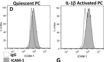
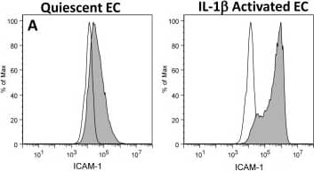
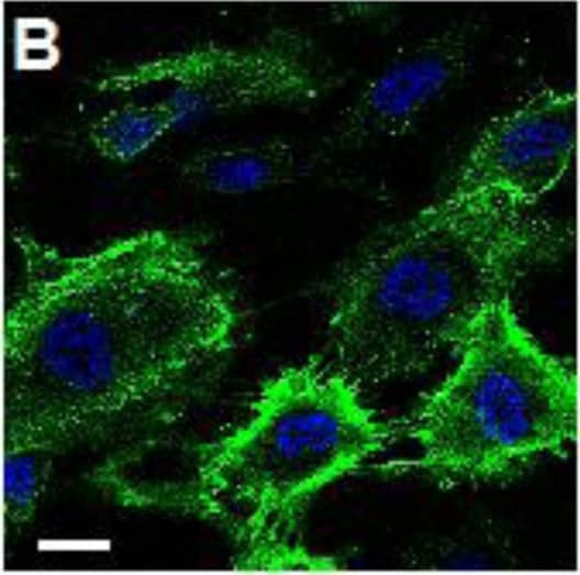
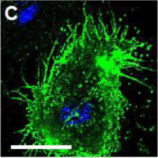
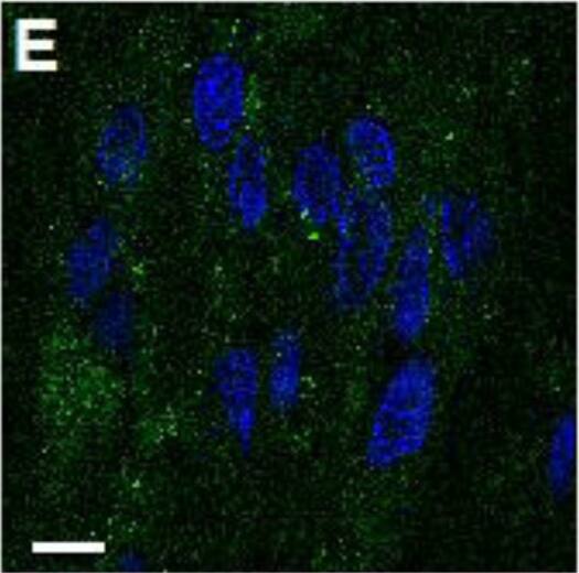
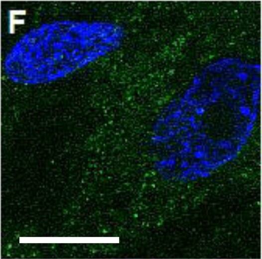
EC and PC ICAM-1 expression and effects of CD18 blocking on neutrophil adhesion.ICAM-1 expression on (A) EC or (D) PC monolayers before and after 4 hr IL-1 beta activation was assessed by flow cytometry (IgG labeled control white). ICAM-1 expression on (B,C) EC and (E,F) PC following 4 hr IL-1 beta activation was imaged using confocal microscopy. Scale bars are 20 µm. (G) Neutrophil adhesion inhibition to IL-1 beta activated EC or PC monolayers. Freshly isolated neutrophils were pre-incubated with anti-Mac-1 or anti-LFA-1 antibodies and seeded onto EC or PC monolayers (activated for 4 with IL-1 beta) in Sykes-Moore chambers and allowed to adhere prior to counting. Bars represent average neutrophil adhesion ± SEM. *P<0.05, when compared to the no block control. Image collected and cropped by CiteAb from the following publication (https://dx.plos.org/10.1371/journal.pone.0060025), licensed under a CC-BY license. Not internally tested by R&D Systems.Detection of Human ICAM-1/CD54 by Flow Cytometry
EC and PC ICAM-1 expression and effects of CD18 blocking on neutrophil adhesion.ICAM-1 expression on (A) EC or (D) PC monolayers before and after 4 hr IL-1 beta activation was assessed by flow cytometry (IgG labeled control white). ICAM-1 expression on (B,C) EC and (E,F) PC following 4 hr IL-1 beta activation was imaged using confocal microscopy. Scale bars are 20 µm. (G) Neutrophil adhesion inhibition to IL-1 beta activated EC or PC monolayers. Freshly isolated neutrophils were pre-incubated with anti-Mac-1 or anti-LFA-1 antibodies and seeded onto EC or PC monolayers (activated for 4 with IL-1 beta) in Sykes-Moore chambers and allowed to adhere prior to counting. Bars represent average neutrophil adhesion ± SEM. *P<0.05, when compared to the no block control. Image collected and cropped by CiteAb from the following publication (https://dx.plos.org/10.1371/journal.pone.0060025), licensed under a CC-BY license. Not internally tested by R&D Systems.Applications for Human ICAM-1/CD54 Fluorescein-conjugated Antibody
Application
Recommended Usage
Flow Cytometry
10 µL/106 cells
Sample: Human peripheral blood mononuclear cells (PBMCs)
Sample: Human peripheral blood mononuclear cells (PBMCs)
Reviewed Applications
Read 3 reviews rated 4.7 using BBA20 in the following applications:
Formulation, Preparation, and Storage
Purification
Protein A or G purified from hybridoma culture supernatant
Formulation
Supplied in a saline solution containing BSA and Sodium Azide.
Shipping
The product is shipped with polar packs. Upon receipt, store it immediately at the temperature recommended below.
Stability & Storage
Protect from light. Do not freeze.
- 12 months from date of receipt, 2 to 8 °C as supplied.
Background: ICAM-1/CD54
Long Name
Intercellular Adhesion Molecule 1
Alternate Names
CD54, ICAM1
Gene Symbol
ICAM1
Additional ICAM-1/CD54 Products
Product Documents for Human ICAM-1/CD54 Fluorescein-conjugated Antibody
Product Specific Notices for Human ICAM-1/CD54 Fluorescein-conjugated Antibody
For research use only
Loading...
Loading...
Loading...
Loading...
Loading...
