Human IL-18 R beta/IL-1 R7 Antibody
R&D Systems, part of Bio-Techne | Catalog # MAB1181

Key Product Details
Validated by
Species Reactivity
Validated:
Cited:
Applications
Validated:
Cited:
Label
Antibody Source
Product Specifications
Immunogen
Met1-Arg356
Accession # O95256
Specificity
Clonality
Host
Isotype
Scientific Data Images for Human IL-18 R beta/IL-1 R7 Antibody
IFN-gamma secretion Induced by IL-18/IL1F4 and Neutralization by Human IL-18 R beta/IL-1 R7 Antibody.
In the presence of Recombinant Human TNF-alpha (20 ng/mL, Catalog # 210-TA), Recombinant Human IL-18/ IL-1F4 stimulates IFN-gamma secretion in the KG-1 human acute myelogenous leukemia cell line in a dose-dependent manner (left-hand graph), as measured by the Human IFN-gamma Quantikine ELISA Kit (Catalog # DIF50C). Under these conditions, IFN-gamma secretion elicited by Recombinant Human IL-18/ IL-1F4 (40 ng/mL) is neutralized (right-hand graph) by increasing concentrations of Mouse Anti-Human IL-18 R beta/IL-1 R7 Monoclonal Antibody (Catalog # MAB1181). The ND50 is typically 0.3-1.0 µg/mL.Detection of Human IL-18 R beta/IL-1 R7/ACPL by Western Blot
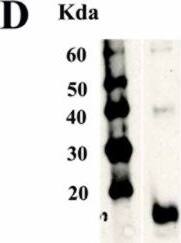
Soluble interleukin-18 receptor alpha complex is associated with interleukin-18 and the soluble form of the interleukin-18 receptor beta chain. (A) Sodium dodecyl sulfate (SDS)-polyacrylamide gel electrophoresis of the serum interleukin (IL)-18 receptor alpha (IL-18R alpha) complex. Purified H44 monoclonal antibody (mAb) was coupled to a 5-mL HiTrap NHS-activated HP column (GE Healthcare). Pooled human blood serum (120 mL) was applied to this affinity column. The IL-18R alpha complex was eluted with elution buffer at a flow rate of 2 mL/minute. Every 1 mL of the elution buffer was collected into a test tube containing 50 mL of neutralization buffer (collected fractions were denoted in order as fractions 1, 2, 3 and so on). A 10-μL aliquot of every fraction (fractions 1 to 8) was treated with the same volume of sample buffer containing 4% SDS (Tris-Glycine SDS Sample Buffer (2×); Invitrogen). Electrophoresis was carried out in the presence of 0.1% SDS, and the gel was stained with Coomassie Brilliant Blue. (B) Western blots of the serum IL-18R alpha complex using an antihuman IL-18R alpha mAb are shown. Western blot analysis was performed using antihuman IL-18R alpha mAb 70625 (R&D Systems, Inc.). Lane 1: Western blot showing 1 μg of rhIL-18R alpha/Fc chimera protein (R&D Systems, Inc.). Lane 2: Western blot showing 5 μg of isolated serum IL-18R alpha complex. (C) Western blot showing serum IL-18R alpha complex using an antihuman IL-18 mAb. Western blot analysis was performed using antihuman IL-18 mAb clone 8 with 5 μg of isolated serum IL-18R alpha complex. (D) Western blot showing serum IL-18R alpha complex using an antihuman IL-18R beta mAb. Western blot analysis was performed using antihuman IL-18R beta mAb 132016 (R&D Systems, Inc.) with 5 μg of isolated serum IL-18R alpha complex. Image collected and cropped by CiteAb from the following publication (https://pubmed.ncbi.nlm.nih.gov/21435242), licensed under a CC-BY license. Not internally tested by R&D Systems.Applications for Human IL-18 R beta/IL-1 R7 Antibody
Western Blot
Sample: Recombinant Human IL-18 R beta/IL-1 R7 Fc Chimera (Catalog # 118-AP)
Neutralization
Formulation, Preparation, and Storage
Purification
Reconstitution
Formulation
Shipping
Stability & Storage
- 12 months from date of receipt, -20 to -70 °C as supplied.
- 1 month, 2 to 8 °C under sterile conditions after reconstitution.
- 6 months, -20 to -70 °C under sterile conditions after reconstitution.
Background: IL-18 R beta/IL-1 R7
IL-18, originally described as an interferon-gamma inducing factor (IGIF), is a member of the IL-1 family of cytokines that has multiple immunoregulatory functions. It has potent IFN-gamma inducing activities and plays a key role in the activation of T helper type 1 (Th1) responses. The functional IL-18 receptor complex consists of two components, the IL-18 R alpha (IL-1 R5) and IL-18 R beta (also termed IL-1 R7 and AcPL) subunits. Both subunits are members of the IL-1 receptor superfamily. Although
IL‑18 R alpha by itself binds IL-18 with low-affinity and IL-18 R beta does not bind IL-18 in vitro, co-expression of IL-18 R alpha and IL-18 R beta is required for high-affinity binding and IL-18 responsiveness. Human IL-18 R beta cDNA encodes a 599 amino acid (aa) residue precursor type I membrane protein with a 14 aa signal peptide, a 342 aa extracellular region containing three immunoglobulin-like domains, a single transmembrane domain and a 222 aa cytoplasmic domain. Human and mouse IL-18 R beta share 65% aa sequence identity. The expression of IL-18 R beta parallels that of IL-18 R alpha and is detected in numerous tissues including lung, spleen, leukocytes and colon.
References
- Born, T.L. et al. (1998) J. Biol. Chem. 273:29445.
- Okamura, H. et al. (2000) in Cytokine Reference, Vol. 2:1605, Academic Press.
Long Name
Alternate Names
Gene Symbol
UniProt
Additional IL-18 R beta/IL-1 R7 Products
Product Documents for Human IL-18 R beta/IL-1 R7 Antibody
Product Specific Notices for Human IL-18 R beta/IL-1 R7 Antibody
For research use only

