Human IL-2 Antibody
R&D Systems, part of Bio-Techne | Catalog # MAB103561


Key Product Details
Validated by
Species Reactivity
Applications
Label
Antibody Source
Product Specifications
Immunogen
Ala21-Thr153
Accession # P60568
Specificity
Clonality
Host
Isotype
Scientific Data Images for Human IL-2 Antibody
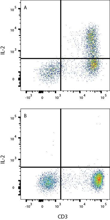
Detection of IL-2 in PBMC cells by Flow Cytometry.
PBMC stimulated with 1 ug/ml of aCD3 and 3 ug/ml of aCD28 for 2 days were treated with Tocris cell activation cocktail 500x (5476) and Brefeldin A (1231/5) for 3 hours (A) or naïve PBMC lymphocytes (B) were stained with Mouse Anti-Human IL-2 Monoclonal Antibody (Catalog # MAB103561) and Mouse Anti-Human CD3 epsilon PE-conjugated Monoclonal Antibody (Catalog # FAB100P).To facilitate intracellular staining, cells were fixed and permeabilized with Flow Cytometry Fixation Buffer (Catalog # FC004). View our protocol for Staining Intracellular Molecules.Applications for Human IL-2 Antibody
Intracellular Staining by Flow Cytometry
Sample: PBMC stimulated with 1 μg/mL of aCD3 and 3 ug/ml of aCD28 for 2 days were treated with Tocris cell activation cocktail 500x (Catalog # 5476) and Brefeldin A (Catalog # 1231/5) for 3 hours or naïve PBMC lymphocytes.
Formulation, Preparation, and Storage
Purification
Reconstitution
Formulation
Shipping
Stability & Storage
- 12 months from date of receipt, -20 to -70 °C as supplied.
- 1 month, 2 to 8 °C under sterile conditions after reconstitution.
- 6 months, -20 to -70 °C under sterile conditions after reconstitution.
Background: IL-2
Recombinant Interleukin-2 (IL-2) is expressed in E. coli and has been engineered to contain the serine for cysteine substitution found in Proleukin® (aldesleukin). Recombinant IL-2 is widely used in cell culture for the expansion of T cells. IL-2 is expressed by CD4+ and CD8+ T cells, gamma delta T cells, B cells, dendritic cells, and eosinophils (1-3). Mature human IL-2 shares 56% and 66% amino acid (aa) sequence identity with mouse and rat IL-2, respectively. Human and mouse IL-2 exhibit cross-species activity (4). The receptor for IL-2 consists of three subunits that are present on the cell surface in varying preformed complexes (5-7). The 55 kDa IL-2 R alpha is specific for IL-2 and binds with low affinity. The 75 kDa IL-2 R beta, which is also a component of the IL-15 receptor, binds IL-2 with intermediate affinity. The 64 kDa common gamma chain gammac/IL-2 R gamma, which is shared with the receptors for IL-4, -7, -9, -15, and -21, does not independently interact with IL-2. Upon ligand binding, signal transduction is performed by both IL-2 R beta and gammac.
IL-2 is best known for its autocrine and paracrine activity on T cells. It drives resting T cells to proliferate and induces IL-2 and IL-2 R alpha synthesis (1, 2). It contributes to T cell homeostasis by promoting the Fas-induced death of naïve CD4+ T cells but not activated CD4+ memory lymphocytes (8). IL-2 plays a central role in the expansion and maintenance of regulatory T cells, although it inhibits the development of Th17 polarized cells (9-11). Thus, IL-2 may be a key cytokine in the natural suppression of autoimmunity (12, 13).
IL-2 expression and concentration can have either immunostimulatory effects at high doses or immunosuppressive effects at low doses due to its preferential binding to different receptor subunits expressed by various immune cell types. This has led to the generation of recombinant IL-2 variants aimed at modifying IL-2 receptor binding for increased antitumor efficacy (14, 15). These variants are typically used in combination with immune checkpoint inhibitors instead of as a monotherapy (14). IL-2 can be genetically engineered to express in NK cells for CAR T cell therapies, and in combination with other cytokines like IL-15, can increase cell viability and proliferation (16). In addition to adoptive cell transfer and checkpoint blockade inhibitors, cancer vaccines that boost immune responses have been combined with IL-2 treatment with promising results in recent studies (15).
In cell culture, IL-2 is a frequently used cytokine for the proliferation, differentiation, and increased antibody secretion of B cells as they transform into plasma cells in vitro (17). IL-2 is also a classically used cytokine for the expansion of NK cells, early differentiated T cells and effector memory Treg cells for adoptive cell transfer cancer immunotherapy (16, 18). GMP IL-2 is a commonly used supplement for the expansion of these cell types for cellular therapies.
References
- Ma, A. et al. (2006) Annu. Rev. Immunol. 24:657.
- Gaffen, S.L. and K.D. Liu (2004) Cytokine 28:109.
- Taniguchi, T. et al. (1983) Nature 302:305.
- Mosmann, T.R. et al. (1987) J. Immunol. 138:1813.
- Liparoto, S.F. et al. (2002) Biochemistry 41:2543.
- Wang, X. et al. (2005) Science 310:1159.
- Bodnar, A. et al. (2008) Immunol. Lett. 116:117.
- Jaleco, S. et al. (2003) J. Immunol. 171:61.
- Malek, T.R. (2003) J. Leukoc. Biol. 74:961.
- Laurence, A. et al. (2007) Immunity 26:371.
- Kryczek, I. et al. (2007) J. Immunol. 178:6730.
- Afzali, B. et al. (2007) Clin. Exp. Immunol. 148:32.
- Fehervari, Z.et al. (2006) Trends Immunol. 27:109.
- Xue, D. et al. (2021) Antibody Therapeutics. 4(2): 123-133.
- Wolfarth, A.A. et al. (2022) Immune Netw. 22(1): e5.
- Koehl, U. et al. (2015) Oncoimmunology. 5(4).
- Marsman, C. et al. (2022) Front. In Immunol. 13(815449).
- Chamucero-Millares, J.A. et al. (2021) Cellular Immunology. 360 (104257).
Long Name
Alternate Names
Entrez Gene IDs
Gene Symbol
UniProt
Additional IL-2 Products
Product Documents for Human IL-2 Antibody
Product Specific Notices for Human IL-2 Antibody
For research use only