Human Klotho beta Antibody
R&D Systems, part of Bio-Techne | Catalog # AF5889

Key Product Details
Species Reactivity
Validated:
Cited:
Applications
Validated:
Cited:
Label
Antibody Source
Product Specifications
Immunogen
Phe53-Leu997
Accession # Q86Z14
Specificity
Clonality
Host
Isotype
Scientific Data Images for Human Klotho beta Antibody
Detection of Human Klotho beta by Western Blot.
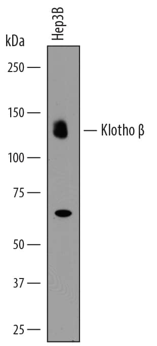
Western blot shows lysates of Hep3B human hepatocellular carcinoma cell line. PVDF membrane was probed with 0.5 µg/mL of Goat Anti-Human Klotho beta Antigen Affinity-purified Polyclonal Antibody (Catalog # AF5889) followed by HRP-conjugated Anti-Goat IgG Secondary Antibody (Catalog # HAF017). A specific band was detected for Klotho beta at approximately 130 kDa (as indicated). This experiment was conducted under reducing conditions and using Immunoblot Buffer Group 1.Klotho beta in HepG2 Human Cell Line.
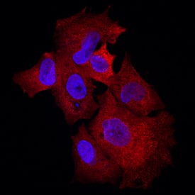
Klotho beta was detected in immersion fixed HepG2 human hepatocellular carcinoma cell line using Goat Anti-Human Klotho beta Antigen Affinity-purified Polyclonal Antibody (Catalog # AF5889) at 10 µg/mL for 3 hours at room temperature. Cells were stained using the NorthernLights™ 557-conjugated Anti-Goat IgG Secondary Antibody (red; Catalog # NL001) and counterstained with DAPI (blue). Specific staining was localized to cytoplasm. View our protocol for Fluorescent ICC Staining of Cells on Coverslips.Applications for Human Klotho beta Antibody
Immunocytochemistry
Sample: Immersion fixed HepG2 human hepatocellular carcinoma cell line
Western Blot
Sample: Hep3B human hepatocellular carcinoma cell line
Formulation, Preparation, and Storage
Purification
Reconstitution
Formulation
Shipping
Stability & Storage
- 12 months from date of receipt, -20 to -70 °C as supplied.
- 1 month, 2 to 8 °C under sterile conditions after reconstitution.
- 6 months, -20 to -70 °C under sterile conditions after reconstitution.
Background: Klotho beta
Klotho beta, a divergent structural member of the glycosidase I superfamily, is expressed primarily in the liver and pancreas, with lower expression in adipose tissue (1, 2). Like Klotho, Klotho beta facilitates binding between FGF19 subfamily members and their receptors via formation of a ternary complex (3). The Klotho beta mediated interaction of human FGF19 (mouse FGF15) with FGF Receptor 4 in the liver negatively regulates bile acid synthesis by controlling the secretion of two key bile acid synthase genes, cholesterol 7-alpha hydroxylase (Cyp7a1) and sterol 12-alpha hydroxylase (Cyp8b1) (2-5). Klotho beta is also a cofactor for the interaction of FGF21 with FGF Receptor 1c in adipocytes, which allows FGF21 to stimulate GLUT1 expression, upregulating adipocyte insulin-dependent glucose uptake (2-4, 6). The 1043 amino acid (aa) type I transmembrane protein is composed of a 51 aa signal sequence, a 943 aa extracellular domain (ECD) containing two glycosidase-like regions, a 21 aa transmembrane domain, and 28 aa intracellular tail. Since Klotho-related proteins lack critical active site Glu residues present in beta-glycosidases, it was initially unclear whether they were functional enzymes (1, 7). However, glucuronidase activity has since been demonstrated for Klotho, indicating that physiologically relevant enzymatic activity for Klotho beta is also possible (8). The extracellular domain shares 79%, 87%, 87% and 67% identity with mouse, equine, canine and rat Klotho beta, respectively. The low identity with rat reflects aa discordance within rodent ECD.
References
- Mian, I. S. (1998) Blood Cells Mol. Dis. 24:83.
- Kurosu, H. and M. Kuro-o (2009) Mol. Cell. Endocrinol. 299:72.
- Ito, S. et al. (2005) J. Clin. Invest. 115:2202.
- Kurosu, H. et al. (2007) J. Biol. Chem. 282:26687.
- Lin, B. C. et al. (2007) J. Biol. Chem. 282:27277.
- Ogawa, Y. et al. (2007) Proc. Natl. Acad. Sci USA 104:7432.
- Chang, Q. et al. (2005) Science 310:490.
- Goetz, R. et al. (2007) Mol. Cell. Biol. 27:3417.
Alternate Names
Gene Symbol
UniProt
Additional Klotho beta Products
Product Documents for Human Klotho beta Antibody
Product Specific Notices for Human Klotho beta Antibody
For research use only

