Human LAG-3 Antibody
R&D Systems, part of Bio-Techne | Catalog # MAB23197


Key Product Details
Species Reactivity
Applications
Label
Antibody Source
Product Specifications
Immunogen
Leu23-Leu450
Accession # P18627
Specificity
Clonality
Host
Isotype
Scientific Data Images for Human LAG-3 Antibody
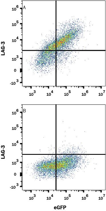
Detection of LAG-3 in HEK293 Human Cell Line Transfected with Human LAG-3 and eGFP by Flow Cytometry.
HEK293 human embryonic kidney cell line transfected with either (A) human LAG-3 or (B) irrelevant transfectants and eGFP was stained with Mouse Anti-Human LAG-3 Monoclonal Antibody (Catalog # MAB23197) followed by APC-conjugated Anti-Mouse IgG Secondary Antibody (Catalog # F0101B). Quadrant markers were set based on control antibody staining (Catalog # MAB0041, data not shown). View our protocol for Staining Membrane-associated Proteins.LAG-3 in Mouse Spleen.
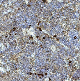
LAG-3 was detected in immersion fixed paraffin-embedded sections of mouse spleen using Mouse Anti-Human LAG-3 Monoclonal Antibody (Catalog # MAB23197) at 15 µg/mL for 1 hour at room temperature followed by incubation with the Anti-Mouse IgG VisUCyte™ HRP Polymer Antibody (VC001). Before incubation with the primary antibody, tissue was subjected to heat-induced epitope retrieval using Antigen Retrieval Reagent-Basic (CTS013). Tissue was stained using DAB (brown) and counterstained with hematoxylin (blue). Specific staining was localized to lymphocytes. Staining was performed using our protocol for IHC Staining with VisUCyte HRP Polymer Detection Reagents.Applications for Human LAG-3 Antibody
CyTOF-ready
Flow Cytometry
Sample: HEK293 Human Cell Line Transfected with Human LAG-3 and eGFP
Immunohistochemistry
Sample: Immersion fixed paraffin-embedded sections of mouse spleen
Formulation, Preparation, and Storage
Purification
Reconstitution
Formulation
Shipping
Stability & Storage
- 12 months from date of receipt, -20 to -70 °C as supplied.
- 1 month, 2 to 8 °C under sterile conditions after reconstitution.
- 6 months, -20 to -70 °C under sterile conditions after reconstitution.
Background: LAG-3
LAG-3 is an activation-induced molecule, expressed on activated T cells and NK cells, but not on resting T cells. Studies using LAG-3 -/- mice have shown significant delay of T cell apoptosis following antigen stimulation and increased size of memory T cells pool following infection (3, 4). It also has been reported that anti-LAG-3 antibodies up-regulate T cell activation by blocking interaction of LAG-3 and MHC class II. The study has demonstrated that LAG-3 is selectively expressed on activated CD4+CD25+ TReg cells and plays a role in their suppressive activity (5). This evidence indicated, unlike the interaction of CD4 with MHC class II that plays a positive role in T cell activation, LAG-3 binds to MHC class II and negatively regulates T cell activation through LAG-3 signaling. On the other hand, studies have shown that binding of LAG-3 to MHC class II molecules on antigen presenting cells induce maturation of dendritic cells and cytokine secretion by monocytes through MHC class II signal transduction (6). Taken together, LAG-3 may have two major functions, it negatively regulates T cells activation through LAG-3 signaling and stimulates antigen presenting cells which express MHC class II.
References
- Triebel, F. et al. (1990) J. Exp. Med. 171:1393.
- Baixeras, E. et al. (1992) J. Exp. Med 176:327.
- Workman, C.J. and D.A. Vignali (2003) Eur. J. Immunol. 33:970.
- Workman, C.J. et al. (2004) J. Immunol. 172:5450.
- Huang, C.T. et al. (2004) Immunity 21:503.
- Andreae, S. et al. (2003) Blood 102:2130.
Long Name
Alternate Names
Gene Symbol
UniProt
Additional LAG-3 Products
Product Documents for Human LAG-3 Antibody
Product Specific Notices for Human LAG-3 Antibody
For research use only
