Human LIGHT/TNFSF14 Antibody
R&D Systems, part of Bio-Techne | Catalog # MAB6643


Key Product Details
Validated by
Species Reactivity
Applications
Label
Antibody Source
Product Specifications
Immunogen
Asp74-Val240
Accession # O43557
Specificity
Clonality
Host
Isotype
Endotoxin Level
Scientific Data Images for Human LIGHT/TNFSF14 Antibody
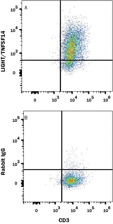
Detection of LIGHT/TNFSF14 in Human CD3+ T cells by Flow Cytometry.
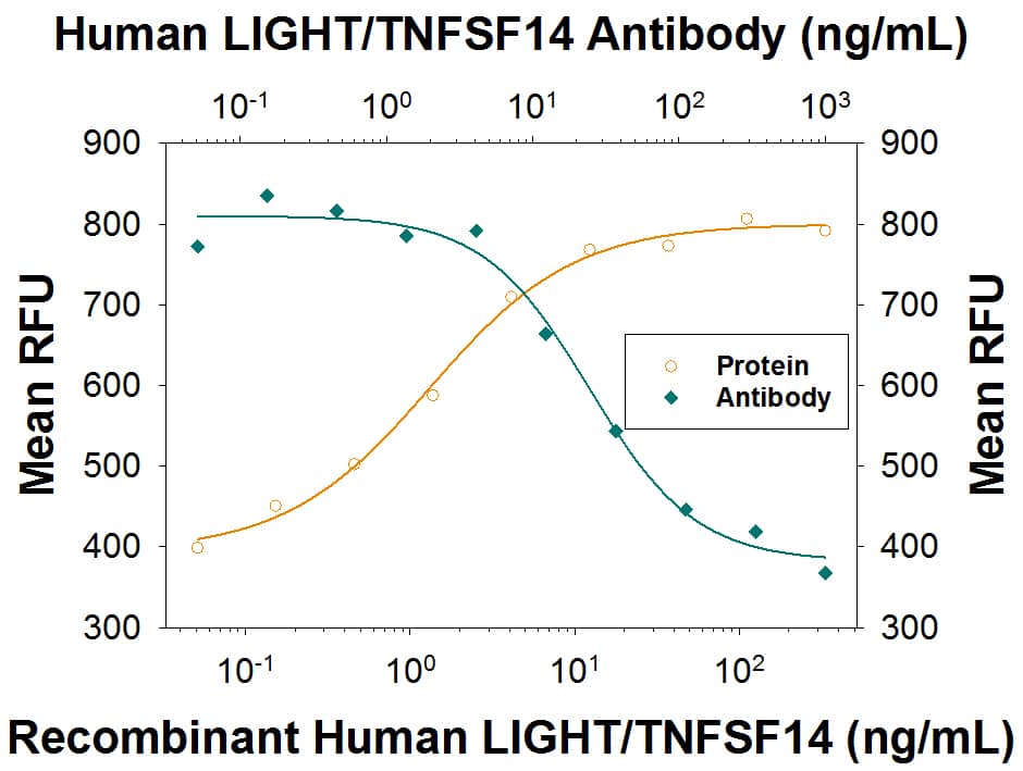
Human CD3+ T cells treated with 1 ng/mL PMA and 1 µg/mL Calcium Ionomycin for 72 hours were stained with (A) Rabbit Anti-Human LIGHT/TNFSF14 Monoclonal Antibody (Catalog # MAB6643) or (B) isotype control antibody (Catalog # MAB1050) followed by anti-Rabbit IgG APC-conjugated secondary antibody (Catalog # F0111) and Mouse Anti-Human CD3 PE-conjugated Monoclonal Antibody (Catalog # FAB100P). View our protocol for Staining Membrane-associated Proteins.Cell Proliferation Induced by LIGHT/TNFSF14 and Neutralization by Human LIGHT/TNFSF14 Antibody.
Recombinant Human LIGHT/TNFSF14 (Catalog # 664-LI) induces proliferation in the HUVEC human umbilical vein endothelial cells in a dose-dependent manner (orange line), as measured by Resazurin (Catalog # AR002). Proliferation elicited by Recombinant Human LIGHT/TNFSF14 (20 ng/mL) is neutralized (green line) by increasing concentrations of Rabbit Anti-Human LIGHT/TNFSF14 Monoclonal Antibody (Catalog # MAB6643). The ND50 is typically 6-48 ng/mL.Applications for Human LIGHT/TNFSF14 Antibody
CyTOF-ready
Flow Cytometry
Sample: Human CD3+ T cells treated with PMA and Calcium Ionomycin
Neutralization
Formulation, Preparation, and Storage
Purification
Reconstitution
Formulation
*Small pack size (-SP) is supplied either lyophilized or as a 0.2 µm filtered solution in PBS.
Shipping
Stability & Storage
- 12 months from date of receipt, -20 to -70 °C as supplied.
- 1 month, 2 to 8 °C under sterile conditions after reconstitution.
- 6 months, -20 to -70 °C under sterile conditions after reconstitution.
Background: LIGHT/TNFSF14
Human LIGHT, also known as TNFSF14, is a type II membrane protein that is a member of the TNF superfamily. LIGHT is an acronym which stands for "is homologous to lymphotoxins, exhibits inducible expression, and competes with HSV glycoprotein D for HVEM, a receptor expressed by T lymphocytes". LIGHT has also been called HVEM-L and LT-gamma. LIGHT is a 240 amino acid (aa) protein that contains a 37 aa cytoplasmic domain, a 22 aa transmembrane region, and a 181 aa extracellular domain. Similar to other TNF ligand family members, LIGHT is predicted to assemble as a homotrimer. LIGHT is produced by activated T cells and was first identified by its ability to compete with HSV glycoprotein D for HVEM binding. LIGHT has also been shown to bind to the lymphotoxin beta receptor (LT betaR) and the decoy receptor (DcR3/TR6). LIGHT overexpression in tumor cells induces apoptosis, which can be enhanced by IFN-gamma.
References
- Mauri, D.N. et al. (1998) Immunity 8:21.
- Zhai, Y. et al. (1998) J. Clin. Invest. 102:1142.
- Harrop, J.A. et al. (1998) J. Biol. Chem. 273:27548.
- Yu, K-Y. et al. (1999) J. Biol. Chem. 274:13733.
Long Name
Alternate Names
Gene Symbol
UniProt
Additional LIGHT/TNFSF14 Products
Product Documents for Human LIGHT/TNFSF14 Antibody
Product Specific Notices for Human LIGHT/TNFSF14 Antibody
For research use only
