Human M-Cadherin/Cadherin-15 Antibody
R&D Systems, part of Bio-Techne | Catalog # AF4096

Key Product Details
Species Reactivity
Validated:
Cited:
Applications
Validated:
Cited:
Label
Antibody Source
Product Specifications
Immunogen
Val22-Ala606
Accession # P55291
Specificity
Clonality
Host
Isotype
Scientific Data Images for Human M-Cadherin/Cadherin-15 Antibody
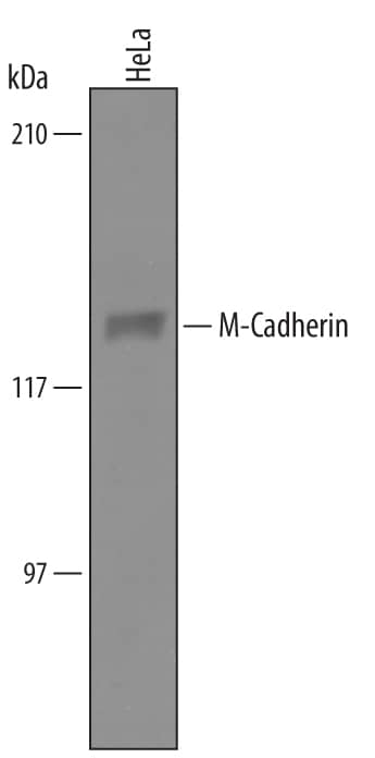
Detection of Human M-Cadherin/Cadherin-15 by Western Blot.
Western blot shows lysates of HeLa human cervical epithelial carcinoma cell line. PVDF Membrane was probed with 1 µg/mL of Sheep Anti-Human M-Cadherin/Cadherin-15 Antigen Affinity-purified Polyclonal Antibody (Catalog # AF4096) followed by HRP-conjugated Anti-Sheep IgG Secondary Antibody (Catalog # HAF016). A specific band was detected for M-Cadherin/Cadherin-15 at approximately 130 kDa (as indicated). This experiment was conducted under reducing conditions and using Immunoblot Buffer Group 8.Detection of M‑Cadherin/ Cadherin‑15 in C2C12 Mouse Cell Line by Flow Cytometry.
C2C12 mouse myoblast cell line was stained with Sheep Anti-Human M-Cadherin/ Cadherin-15 Antigen Affinity-purified Polyclonal Antibody (Catalog # AF4096, filled histogram) or control antibody (Catalog # 5-001-A, open histogram), followed by NorthernLights™ 557-conjugated Anti-Sheep IgG Secondary Antibody (Catalog # NL010).M‑Cadherin/Cadherin-15 in C2C12 Mouse Cell Line.
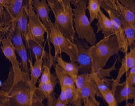
M-Cadherin/Cadherin-15 was detected in immersion fixed C2C12 mouse myoblast cell line using Sheep Anti-Human M-Cadherin/Cadherin-15 Antigen Affinity-purified Polyclonal Antibody (Catalog # AF4096) at 10 µg/mL for 3 hours at room temperature. Cells were stained using the Northern-Lights™ 557-conjugated Anti-Mouse IgG Secondary Antibody (red; Catalog # NL007) and counter-stained with DAPI (blue). Specific staining was localized to cell surfaces and cytoplasm. View our protocol for Fluorescent ICC Staining of Cells on Coverslips.Applications for Human M-Cadherin/Cadherin-15 Antibody
CyTOF-ready
Flow Cytometry
Sample: C2C12 mouse myoblast cell line
Immunocytochemistry
Sample: Immersion fixed C2C12 mouse myoblast cell line
Western Blot
Sample: HeLa human cervical epithelial carcinoma cell line
Formulation, Preparation, and Storage
Purification
Reconstitution
Formulation
Shipping
Stability & Storage
- 12 months from date of receipt, -20 to -70 °C as supplied.
- 1 month, 2 to 8 °C under sterile conditions after reconstitution.
- 6 months, -20 to -70 °C under sterile conditions after reconstitution.
Background: M-Cadherin/Cadherin-15
M-Cadherin (M-CAD or Cadherin-15) is a 124 kDa type I transmembrane glycoprotein of the Cadherin superfamily of calcium-dependent homotypic adhesion molecules (1-4). Like other classical Cadherins, the 814 amino acid (aa) human M-CAD contains a signal sequence (21 aa), a propeptide (29 aa), an extracellular domain with five Cadherin domain repeats (ECD, 556 aa), a transmembrane segment (20 aa) and a cytoplasmic domain (188 aa) (1). A splice variant that diverges within the third Cadherin repeat has been sequenced. The Cadherin repeats are responsible for cell-cell adhesion by homophilic binding on opposing cells (1-4). Intracellularly, M-CAD binds beta-catenin or plakoglobin ( gamma-catenin), which in turn bind alpha-catenin (4). M-CAD also binds p120 catenin (5). Connection of Cadherin/catenin complexes to the actin cytoskeleton is in question, but is possible through a linker (6). Connection to microtubules has been shown (7). M-CAD is present during early stages of skeletal muscle development and is thought to align myoblasts for fusion (8). It is also present in muscle satellite cells and participates in muscle regeneration (9). M-CAD is also expressed in the granule cell layer of the cerebellar glomerulus (10). Deletion of mouse M-CAD has little effect in vivo, most likely due to compensation by N-Cadherin (11). However, M-CAD upregulation and adhesion between myoblasts during induction of differentiation in vitro is required for their fusion (8, 12, 13). M-CAD activity is later downregulated by sequestering to caveoli, p120 catenin/RhoA-induced ubiquitination and/or cleavage by calpain-3. This terminates fusion and allows sarcomere formation (5, 12, 13). Human M-CAD ECD shows 88% aa identity with mouse, rat, or bovine and 85% aa identity with canine M-CAD ECD. M-CAD is an outlier among classical Cadherins, with 40% aa identity or less in the ECD (4).
References
- Kaufmann, U. et al. (1999) Cell Tissue Res. 296:191.
- Angst, B.D. et al. (2001) J. Cell Sci. 114:629.
- Shimoyama, Y. et al. (1998) J. Biol. Chem. 273:10011.
- Kuch, C. et al. (1997) Exp. Cell Res. 232:331.
- Charasse, S. et al. (2006) Mol. Biol. Cell 17:749.
- Weis, W.I. and W. J. Nelson (2006) J. Biol. Chem. 281:35593.
- Kaufmann, U. et al. (1999) J. Cell Sci. 112:55.
- Zeschnigk, M. et al. (1995) J. Cell Sci. 108:2973.
- Wrobel, E. et al. (2007) Eur. J. Cell Biol. Jan 10; [Epub ahead of print]
- Rose, O. et al. (1995) Proc. Natl. Acad. Sci. USA 92:6022.
- Hollnagel, A. et al. (2002) Mol. Cell. Biol. 22:4760.
- Volonte, D. et al. (2003) Mol. Biol. Cell 14:4075.
- Kramerova, I. et al. (2006) Mol. Cell. Biol. 26:8437.
Long Name
Alternate Names
Gene Symbol
UniProt
Additional M-Cadherin/Cadherin-15 Products
Product Documents for Human M-Cadherin/Cadherin-15 Antibody
Product Specific Notices for Human M-Cadherin/Cadherin-15 Antibody
For research use only


