Human Matrilin-2 Antibody
R&D Systems, part of Bio-Techne | Catalog # AF3044

Key Product Details
Validated by
Species Reactivity
Applications
Label
Antibody Source
Product Specifications
Immunogen
Arg24-Arg937
Accession # AAH10444
Specificity
Clonality
Host
Isotype
Scientific Data Images for Human Matrilin-2 Antibody
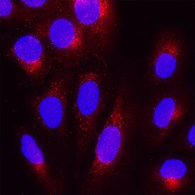
Matrilin-2 in U2OS Human Cell Line.
Matrilin-2 was detected in immersion fixed U2OS human osteosarcoma cell line using Goat Anti-Human Matrilin-2 Antigen Affinity-purified Polyclonal Antibody (Catalog # AF3044) at 5 µg/mL for 3 hours at room temperature. Cells were stained using the NorthernLights™ 557-conjugated Anti-Goat IgG Secondary Antibody (red; NL001) and counterstained with DAPI (blue). Specific staining was localized to cytoplasm. Staining was performed using our protocol for Fluorescent ICC Staining of Non-adherent Cells.Detection of Mouse Matrilin-2 by Immunohistochemistry
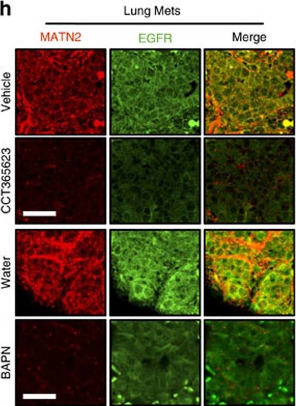
Chemical inhibition of LOX blocks tumour growth and metastasis.(a) Tumour growth in vehicle or CCT365623-treated MMTV-PyMT mice. Data are represented as mean±s.d. from six mice per group. **P<0.01, Student's t-test. (b) H&E stained lungs from vehicle or CCT365623 treated MMTV-PyMT mice. Scale bar, 100 μm. (c) Quantification of lung metastasis in mice from a. All data are presented as mean±range, n=6 animals. **P<0.05, Mann–Whitney analysis. (d) Tumour growth in water or BAPN-treated MMTV-PyMT mice. Data are represented as mean±s.d. from seven mice per group. **P<0.01, Student's t-test. (e) H&E stained lungs from water or BAPN-treated MMTV-PyMT mice. Scale bar, 100 μm. (f) Quantification of lung metastasis in mice from d. All data are presented as mean±range, n=7 animals. **P<0.05, Mann–Whitney analysis. (g,h) Confocal photomicrographs of MATN2 (red) and EGFR (green) staining in primary and metastatic tumours from vehicle, CCT365623, water or BAPN-treated MMTV-PyMT mice. Scale bars, 20 μm. (i–l) Quantification of MATN2 and EGFR staining in primary or metastatic tumours from g and h. All data are represented as mean±s.e.m. Samples from six vehicle or CCT365623 and seven water or BAPN-treated mice were analysed. **P<0.01, Student's t-test. Image collected and cropped by CiteAb from the following publication (https://www.nature.com/articles/ncomms14909), licensed under a CC-BY license. Not internally tested by R&D Systems.Detection of Mouse Matrilin-2 by Immunohistochemistry
LOX controls EGF-dependent cell proliferation in vitro and tumour growth in vivo.(a,b) Graphs showing cell proliferation in control (shCtl) and LOX-depleted (shLOX A,B) MDA-MB-231 (a) and U87 (b) cells grown in 10% FBS (serum) or serum-free medium (serum free) and treated with PBS, rhMATN2 (500 ng ml−1) and EGF (50 ng ml−1) as indicated. All data are represented as mean±s.d. from four independent experiments. **P<0.01, Student's t-test. (c) Kaplan–Meier survival plot for mice following tail vein injection of control (shCtl) or LOX-depleted (shLOX A) MDA-MB-231 cells. Seven mice were used in each group. (d) H&E staining of lungs from mice in c. Scale bar, 500 μm. (e) Quantification of lung deposits in mice. Data is represented as mean±s.d. from seven samples. **P<0.05, Mann–Whitney analysis. (f) Photomicrographs for MATN2 (red) and EGFR (green) in lung deposits. Scale bar, 10 μm. (g) Quantification of samples in f. Data is represented as mean±s.e.m. from six samples. **P<0.01, Student's t-test. Image collected and cropped by CiteAb from the following publication (https://www.nature.com/articles/ncomms14909), licensed under a CC-BY license. Not internally tested by R&D Systems.Applications for Human Matrilin-2 Antibody
Immunocytochemistry
Sample: Immersion fixed U2OS human osteosarcoma cell line
Western Blot
Sample: Recombinant Human Matrilin-2 (Catalog # 3044-MN)
Formulation, Preparation, and Storage
Purification
Reconstitution
Formulation
Shipping
Stability & Storage
- 12 months from date of receipt, -20 to -70 °C as supplied.
- 1 month, 2 to 8 °C under sterile conditions after reconstitution.
- 6 months, -20 to -70 °C under sterile conditions after reconstitution.
Background: Matrilin-2
Matrilin-2 is an extracellular matrix protein that belongs to the superfamily of von Willebrand factor A (VWA) containing proteins. It is expressed in many tissues and functions as a bridging component between other matrix proteins (1‑4). The human Matrilin-2 cDNA encodes a 956 amino acid (aa) precursor with a 23 aa signal sequence, two VWA domains separated by ten tandem EGF-like repeats, and a C-terminal coiled-coil domain (5, 6). Alternate splicing generates Isoform 2 (with an 18 aa deletion near the C-terminus), Isoform 3 (with a deletion of the fourth EGF-like repeat), and Isoform 4 (with a deletion of the first VWA and first EGF-like repeat). Human Matrilin-2 shares 87% and 84% aa sequence identity with mouse and canine Matrilin-2, respectively, and 27%, 22%, and 33% aa sequence identity with human Matrilin-1, -3, and -4, respectively. Matrilin-2 forms a variety of disulfide-linked oligomers via its coiled-coil domain (4, 7, 8, 9). It can assemble into homotrimers or heterotrimers with Matrilin-1 and/or Matrilin-4 (4, 7, 8) but has not been detected in heterotrimers containing Matrilin-3 (8). The VWA domains are thought to mediate Matrilin-Matrilin interactions as well as interactions with other matrix proteins such as Fibronectin, Collagen I, Fibrillin-2, and Laminin-1/Nidogen-1 complexes (7). Matrilin-2 knockout mice do not display any obvious abnormalities, suggesting that the expression of other molecules can compensate for the lack of Matrilin-2 (10).
References
- Wagener, R. et al. (2005) FEBS Lett. 579:3323.
- Deak, F. et al. (1999) Matrix Biol. 18:55.
- Whittaker, C.A. and R.O. Hynes (2002) Mol. Biol. Cell 13:3369.
- Piecha, D. et al. (1999) J. Biol. Chem. 274:13353.
- Muratoglu, S. et al. (2000) Cytogenet. Cell Genet. 90:323.
- Deak, F. et al. (1997) J. Biol. Chem. 272:9268.
- Piecha, D. et al. (2002) Biochem. J. 367:715.
- Frank, S. et al. (2002) J. Biol. Chem. 277:19071.
- Pan, O.H. and K. Beck (1998) J. Biol. Chem. 273:14205.
- Mates, L. et al. (2004) Matrix Biol. 23:195.
Alternate Names
Entrez Gene IDs
Gene Symbol
UniProt
Additional Matrilin-2 Products
Product Documents for Human Matrilin-2 Antibody
Product Specific Notices for Human Matrilin-2 Antibody
For research use only


